Copyright
©2008 The WJG Press and Baishideng.
World J Gastroenterol. Jul 14, 2008; 14(26): 4196-4203
Published online Jul 14, 2008. doi: 10.3748/wjg.14.4196
Published online Jul 14, 2008. doi: 10.3748/wjg.14.4196
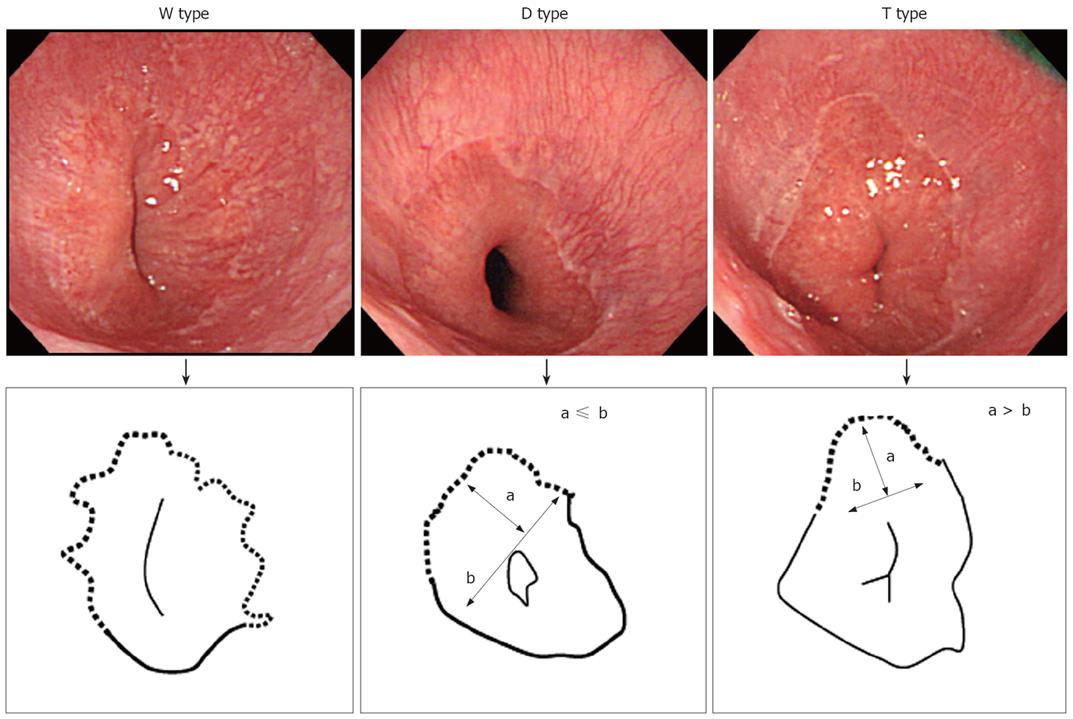
Figure 1 The shape of ESEM.
We originally divided ESEM into 3 types based on its shape. The W type, in which the ESEM was defined by a columnar epithelium which extended continuously from the gastric lumen to the esophagus, in which the uppermost extent of the visible red columnar epithelium could be observed as a wave-like formation (W). The broken line in the illustration shows the ESEM. The D type, in which the length of basal part of the ESEM (b) was longer than the length of the major part of the ESEM (a; i.e. a < b) The columnar epithelium of the ESEM is observed as a dome-like shape (D). The broken line in the illustration shows the ESEM. The T type in which the length of the major axis of the ESEM (a) is longer than the basal part of ESEM (b; i.e. a ≥ b). The columnar epithelium of the ESEM is observed as a tongue-like shape (T).
- Citation: Yamagishi H, Koike T, Ohara S, Kobayashi S, Ariizumi K, Abe Y, Iijima K, Imatani A, Inomata Y, Kato K, Shibuya D, Aida S, Shimosegawa T. Tongue-like Barrett’s esophagus is associated with gastroesophageal reflux disease. World J Gastroenterol 2008; 14(26): 4196-4203
- URL: https://www.wjgnet.com/1007-9327/full/v14/i26/4196.htm
- DOI: https://dx.doi.org/10.3748/wjg.14.4196


