Copyright
©2008 The WJG Press and Baishideng.
World J Gastroenterol. Jul 14, 2008; 14(26): 4179-4184
Published online Jul 14, 2008. doi: 10.3748/wjg.14.4179
Published online Jul 14, 2008. doi: 10.3748/wjg.14.4179
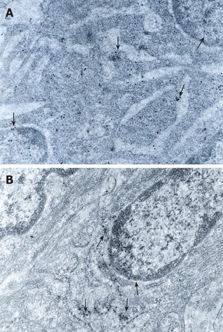
Figure 4 A: Electron micrograph from a mild-grade dysplasia adenoma showing GST-pi positive particles located in ribosomes and nucleus (arrows, × 42 000); B: Ultrathin section from a high-grade dysplasia adenoma showing GST-pi positive particles located in cytoplasmic membranes and nuclear membrane (with uranylacetate and lead citrate arrows, × 57 000).
- Citation: Gaitanarou E, Seretis E, Xinopoulos D, Paraskevas E, Arnoyiannaki N, Voloudakis-Baltatzis I. Immunohistochemical localization of glutathione S-transferase-pi in human colorectal polyps. World J Gastroenterol 2008; 14(26): 4179-4184
- URL: https://www.wjgnet.com/1007-9327/full/v14/i26/4179.htm
- DOI: https://dx.doi.org/10.3748/wjg.14.4179


