Copyright
©2008 The WJG Press and Baishideng.
World J Gastroenterol. Jul 14, 2008; 14(26): 4146-4151
Published online Jul 14, 2008. doi: 10.3748/wjg.14.4146
Published online Jul 14, 2008. doi: 10.3748/wjg.14.4146
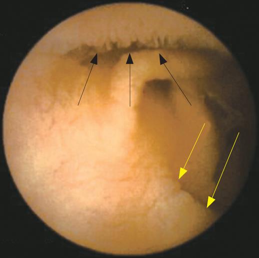
Figure 3 “Patchy” atrophy detected by capsule endoscopy.
Capsule endoscopy shows a normal villous pattern in the upper part of the image (black arrows) and villous atrophy in the lower part (yellow arrows).
- Citation: Spada C, Riccioni ME, Urgesi R, Costamagna G. Capsule endoscopy in celiac disease. World J Gastroenterol 2008; 14(26): 4146-4151
- URL: https://www.wjgnet.com/1007-9327/full/v14/i26/4146.htm
- DOI: https://dx.doi.org/10.3748/wjg.14.4146


