Copyright
©2008 The WJG Press and Baishideng.
World J Gastroenterol. Jul 7, 2008; 14(25): 4093-4095
Published online Jul 7, 2008. doi: 10.3748/wjg.14.4093
Published online Jul 7, 2008. doi: 10.3748/wjg.14.4093
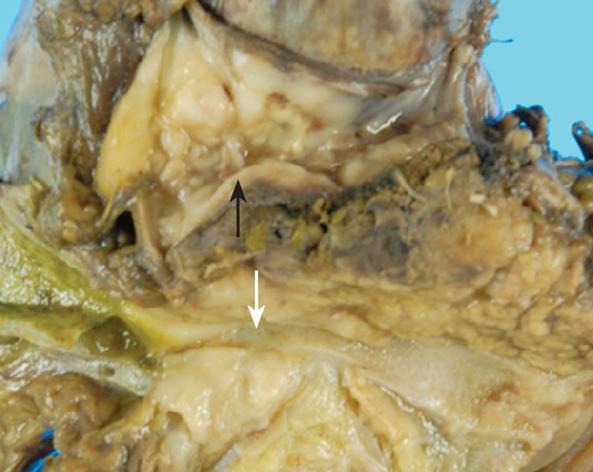
Figure 2 A photograph of the resected specimen.
Endoluminal invasion of both the bile duct (white arrow) and the portal vein (black arrow) can be observed.
- Citation: Hashimoto M, Umekita N, Noda K. Non-Hodgkin lymphoma as a cause of obstructive jaundice with simultaneous extrahepatic portal vein obstruction: A case report. World J Gastroenterol 2008; 14(25): 4093-4095
- URL: https://www.wjgnet.com/1007-9327/full/v14/i25/4093.htm
- DOI: https://dx.doi.org/10.3748/wjg.14.4093


