Copyright
©2008 The WJG Press and Baishideng.
World J Gastroenterol. Jul 7, 2008; 14(25): 4028-4039
Published online Jul 7, 2008. doi: 10.3748/wjg.14.4028
Published online Jul 7, 2008. doi: 10.3748/wjg.14.4028
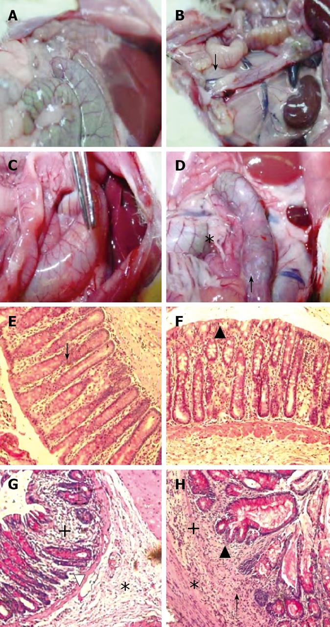
Figure 3 Representative photographs of macroscopic (upper row) and microscopic (lower row, HE stain x 200 ) findings on day 70 in treatment groups MC, B, IA and IA + B.
A and B: Normal colon, note the whitish area at the site of multiple inoculation of the EPEC in colons of B-treated animals (↓); C: Note the vasodilatation and enlargement of the colon (forceps) in the IA treated rats; D: Note adhesions (*), hyperemia and vasodilatation of an enlarged colon and a darkened site reflecting blackish mucosa (↑) in the IA + B treated animals; E and F: Normal microscopic appearance of the colon in the MC and B-treated groups. Well aligned parallel crypts (↓) continuous epithelial lining (▲) normal muscularis mucosa and normal lamina propria infiltration by cells; G: Shows cryptitis (↓), infiltration of inflammatory cells (+) and edema (*) in the submucosal with thinning of the muscularis mucosa and vasodilatation (∆); H: Crypt deformities and bifurcation (▲), cryptitis (↑), extensive inflammatory cells infiltrate (+) and edema (*).
- Citation: Hussein IAH, Tohme R, Barada K, Mostafa MH, Freund JN, Jurjus RA, Karam W, Jurjus A. Inflammatory bowel disease in rats: Bacterial and chemical interaction. World J Gastroenterol 2008; 14(25): 4028-4039
- URL: https://www.wjgnet.com/1007-9327/full/v14/i25/4028.htm
- DOI: https://dx.doi.org/10.3748/wjg.14.4028


