Copyright
©2008 The WJG Press and Baishideng.
World J Gastroenterol. Jul 7, 2008; 14(25): 4005-4010
Published online Jul 7, 2008. doi: 10.3748/wjg.14.4005
Published online Jul 7, 2008. doi: 10.3748/wjg.14.4005
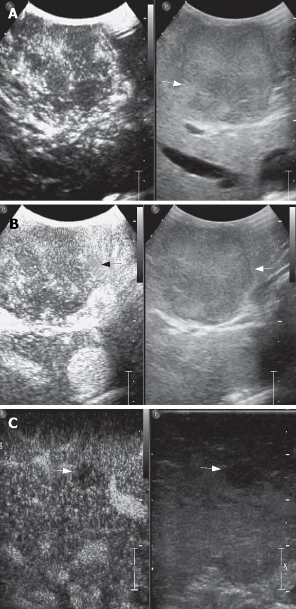
Figure 2 A mosaic nodule in a cirrhotic liver at IOUS.
A: At arterial phase, the nodule shows early enhancement (arrows); B: The nodule demonstrates contrast agent wash-out in late phase: it was a typical appearance of HCC and proved malignant at histology (arrows); C: Another hypoechoic nodule at IOUS was hypoenhanced in late phase: it was considered intrahepatic metastasis and confirmed malignant at histology (arrows).
- Citation: Lu Q, Luo Y, Yuan CX, Zeng Y, Wu H, Lei Z, Zhong Y, Fan YT, Wang HH, Luo Y. Value of contrast-enhanced intraoperative ultrasound for cirrhotic patients with hepatocellular carcinoma: A report of 20 cases. World J Gastroenterol 2008; 14(25): 4005-4010
- URL: https://www.wjgnet.com/1007-9327/full/v14/i25/4005.htm
- DOI: https://dx.doi.org/10.3748/wjg.14.4005


