Copyright
©2008 The WJG Press and Baishideng.
World J Gastroenterol. Jun 28, 2008; 14(24): 3829-3840
Published online Jun 28, 2008. doi: 10.3748/wjg.14.3829
Published online Jun 28, 2008. doi: 10.3748/wjg.14.3829
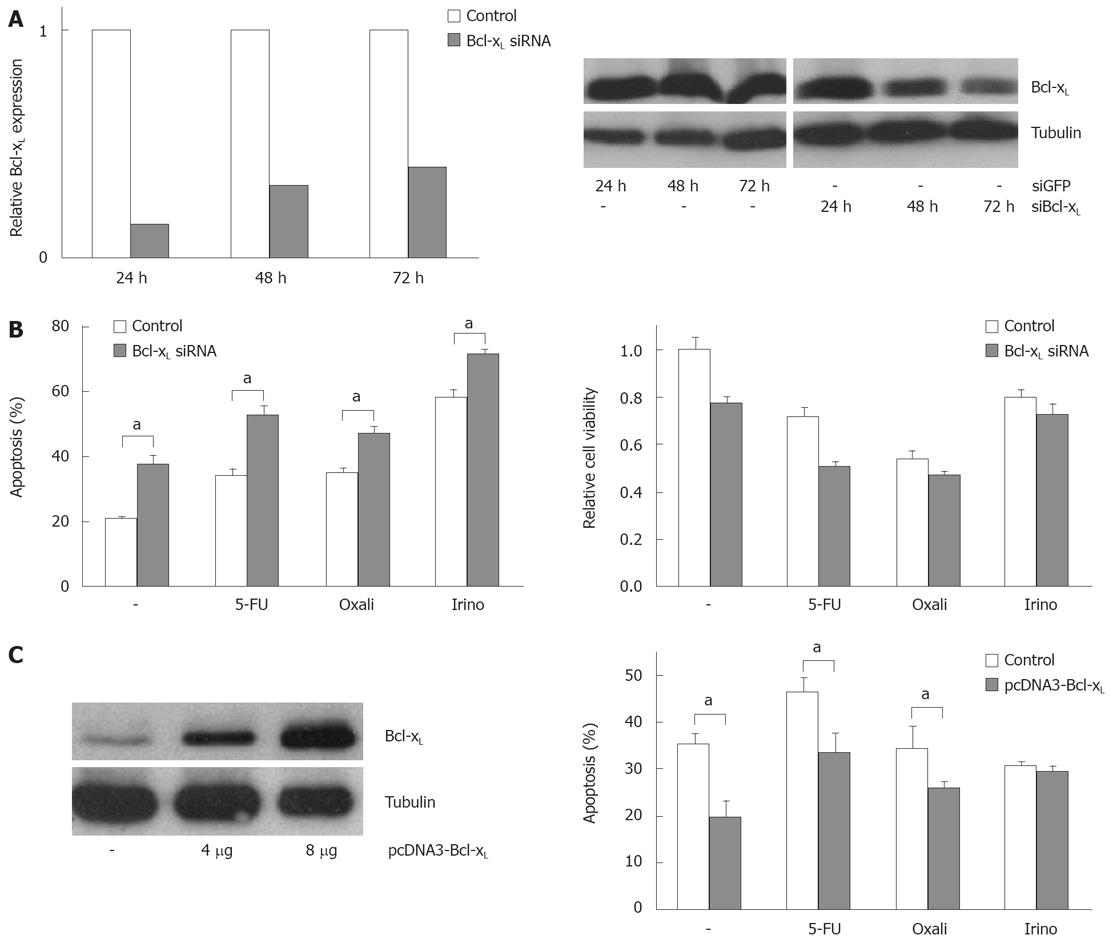
Figure 3 Modulation of Bcl-xL expression alters chemotherapeutic drug-induced apoptosis.
A: SW480 cells were transfected with siRNA specific for Bcl-xL or transfected with siRNA specific for GFP as control (20 nmol/L). After the indicated time post transfection, total RNA was extracted and analyzed for Bcl-xL expression by quantitative real-time PCR (left panel). Relative expression was calculated as described in the materials and methods section. In addition, after the indicated time post transfection, cells were lyzed and analyzed for Bcl-xL expression by Western blot (right panel). α-Tubulin expression was used to control equal loading; B: SW480 cells were transfected with siRNA specific for Bcl-xL or transfected with siRNA specific for GFP as control. 24 h post transfection, cells were treated with 5-FU (10 &mgr;g/mL), oxaliplatin (16 &mgr;g/mL) and irinotecan (60 &mgr;g/mL) for further 24 h. Cells were then harvested and analyzed for apoptosis induction (left panel). In addition, cell viability was measured by MTT assay and is shown relative to mock treated controls (right panel); C: SW480 cells were transfected with pcDNA3 Bcl-xL or pcDNA3 empty vector as control. 24 h post transfection, cells were analyzed for Bcl-xL expression using Western blotting (left panel). In addition, 24 h post transfection, cells were treated with 5-FU (15 &mgr;g/mL), oxaliplatin (10 &mgr;g/mL) and irinotecan (40 &mgr;g/mL) for further 24 h. Cells were then harvested and analyzed for apoptosis induction by flow cytometry. (B) and (C) assays were performed in triplicates. Values are means ± SD, aP < 0.05.
- Citation: Schulze-Bergkamen H, Ehrenberg R, Hickmann L, Vick B, Urbanik T, Schimanski CC, Berger MR, Schad A, Weber A, Heeger S, Galle PR, Moehler M. Bcl-xL and Myeloid cell leukaemia-1 contribute to apoptosis resistance of colorectal cancer cells. World J Gastroenterol 2008; 14(24): 3829-3840
- URL: https://www.wjgnet.com/1007-9327/full/v14/i24/3829.htm
- DOI: https://dx.doi.org/10.3748/wjg.14.3829


