Copyright
©2008 The WJG Press and Baishideng.
World J Gastroenterol. Jun 28, 2008; 14(24): 3829-3840
Published online Jun 28, 2008. doi: 10.3748/wjg.14.3829
Published online Jun 28, 2008. doi: 10.3748/wjg.14.3829
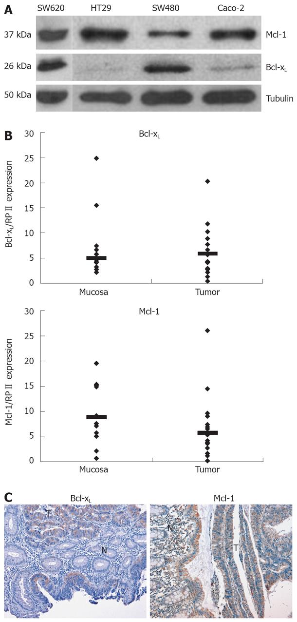
Figure 1 Mcl-1 and Bcl-xL expression in CRC.
A: The CRC cell lines, HT29, SW620, SW480, and Caco-2, were analyzed for the basal expression of the Bcl-2 family members Bcl-xL and Mcl-1. Whole cell lysates were prepared, separated, and immunoblotted with antibodies against Bcl-xL, Mcl-1 and α-tubulin; B: CRC tissues and normal colorectal tissues were tested for mRNA expression (n = 9 patients). mRNA expression levels of Bcl-xL, Mcl-1 and RPII were measured in all tissue samples by quantitative real-time PCR. mRNA expression levels of Bcl-xL or Mcl-1 were normalized to RPIIin each sample. Each PCR reaction was run in triplicates. Median is added; C: Immunohistochemical analysis of human CRC tissues was performed as described in the Methods section. All sections included carcinoma as well as normal epithelial tissues to directly compare Bcl-xL as well as Mcl-1 expression in neoplastic and non-malignant tissues. Representative analysis of immunoperoxidase detection of Bcl-xL and Mcl-1 in paraffin embedded carcinoma tissue (T) and adjacent non-tumor tissue (N) is presented.
- Citation: Schulze-Bergkamen H, Ehrenberg R, Hickmann L, Vick B, Urbanik T, Schimanski CC, Berger MR, Schad A, Weber A, Heeger S, Galle PR, Moehler M. Bcl-xL and Myeloid cell leukaemia-1 contribute to apoptosis resistance of colorectal cancer cells. World J Gastroenterol 2008; 14(24): 3829-3840
- URL: https://www.wjgnet.com/1007-9327/full/v14/i24/3829.htm
- DOI: https://dx.doi.org/10.3748/wjg.14.3829


