Copyright
©2008 The WJG Press and Baishideng.
World J Gastroenterol. Jun 21, 2008; 14(23): 3763-3767
Published online Jun 21, 2008. doi: 10.3748/wjg.14.3763
Published online Jun 21, 2008. doi: 10.3748/wjg.14.3763
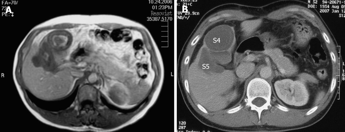
Figure 4 MRI on T1-WI 30 mo after treatment with Imatinib (A) and contrast-enhanced CT 34 mo after the treatment with Imatinib (B).
The metastatic lesions (S4 + S5) are indicated.
- Citation: Suzuki S, Sasajima K, Miyamoto M, Watanabe H, Yokoyama T, Maruyama H, Matsutani T, Liu A, Hosone M, Maeda S, Tajiri T. Pathologic complete response confirmed by surgical resection for liver metastases of gastrointestinal stromal tumor after treatment with imatinib mesylate. World J Gastroenterol 2008; 14(23): 3763-3767
- URL: https://www.wjgnet.com/1007-9327/full/v14/i23/3763.htm
- DOI: https://dx.doi.org/10.3748/wjg.14.3763


