Copyright
©2008 The WJG Press and Baishideng.
World J Gastroenterol. Jun 21, 2008; 14(23): 3616-3620
Published online Jun 21, 2008. doi: 10.3748/wjg.14.3616
Published online Jun 21, 2008. doi: 10.3748/wjg.14.3616
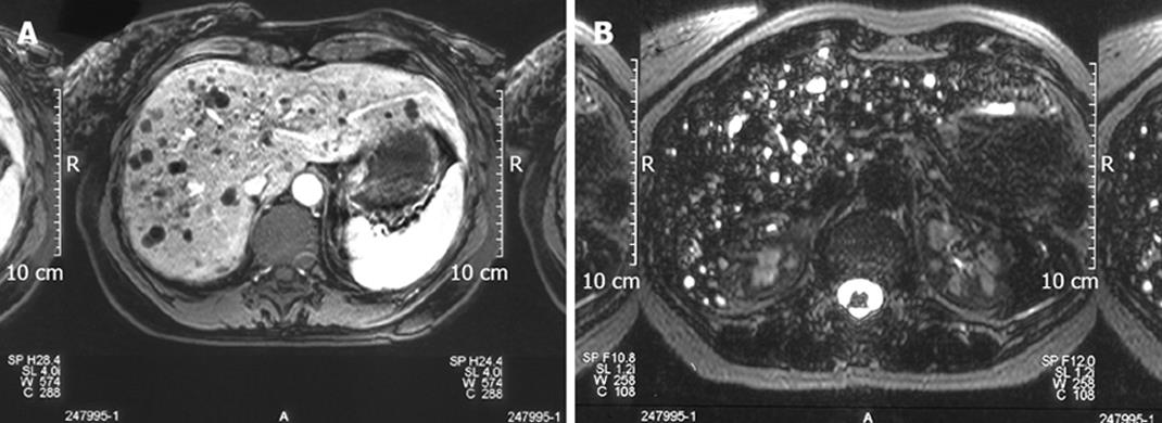
Figure 2 T1 weighted images showing the left multiple hypointense focal lesions of the liver parenchyma simulating metastases (A) and T2 weighted images showing a strong hyper-intensity in Von Meyenburg complex disclosing its cystic nature (B).
- Citation: Poggio PD, Buonocore M. Cystic tumors of the liver: A practical approach. World J Gastroenterol 2008; 14(23): 3616-3620
- URL: https://www.wjgnet.com/1007-9327/full/v14/i23/3616.htm
- DOI: https://dx.doi.org/10.3748/wjg.14.3616


