Copyright
©2008 The WJG Press and Baishideng.
World J Gastroenterol. Jun 14, 2008; 14(22): 3504-3510
Published online Jun 14, 2008. doi: 10.3748/wjg.14.3504
Published online Jun 14, 2008. doi: 10.3748/wjg.14.3504
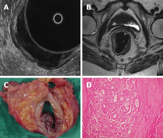
Figure 2 A: ERUS shows the tumor invading the muscularis propria which can be regarded as a T2 tumor.
A lymph node is also seen; B: MRI clearly demonstrates that the tumor is confined to the muscularis propria and does not invade the mesorectal fatty tissue. Lymph nodes are also seen; C: Macroscopic specimen reveals the tumor does not extend to the mesorectal fat; D: Pathology confirms that this is a T2 stage tumor.
-
Citation: Halefoglu AM, Yildirim S, Avlanmis O, Sakiz D, Baykan A. Endorectal ultrasonography
versus phased-array magnetic resonance imaging for preoperative staging of rectal cancer. World J Gastroenterol 2008; 14(22): 3504-3510 - URL: https://www.wjgnet.com/1007-9327/full/v14/i22/3504.htm
- DOI: https://dx.doi.org/10.3748/wjg.14.3504


