Copyright
©2008 The WJG Press and Baishideng.
World J Gastroenterol. Jun 14, 2008; 14(22): 3476-3483
Published online Jun 14, 2008. doi: 10.3748/wjg.14.3476
Published online Jun 14, 2008. doi: 10.3748/wjg.14.3476
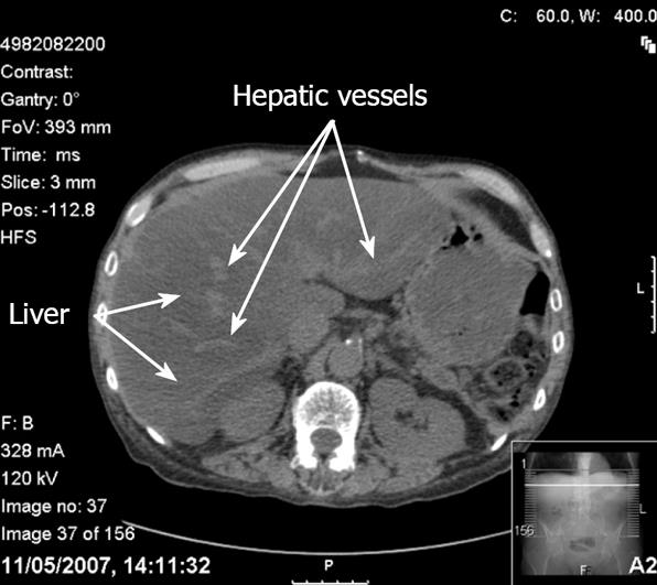
Figure 2 CT findings in hepatic steatosis.
In this contrast unenhanced scan, the liver appears darker than the spleen and the hepatic vessels appear bright. The increased brightness of the vessels relative to the liver parenchyma may erroneously suggest the use of contrast.
- Citation: Mehta SR, Thomas EL, Bell JD, Johnston DG, Taylor-Robinson SD. Non-invasive means of measuring hepatic fat content. World J Gastroenterol 2008; 14(22): 3476-3483
- URL: https://www.wjgnet.com/1007-9327/full/v14/i22/3476.htm
- DOI: https://dx.doi.org/10.3748/wjg.14.3476


