Copyright
©2008 The WJG Press and Baishideng.
World J Gastroenterol. Jan 14, 2008; 14(2): 224-230
Published online Jan 14, 2008. doi: 10.3748/wjg.14.224
Published online Jan 14, 2008. doi: 10.3748/wjg.14.224
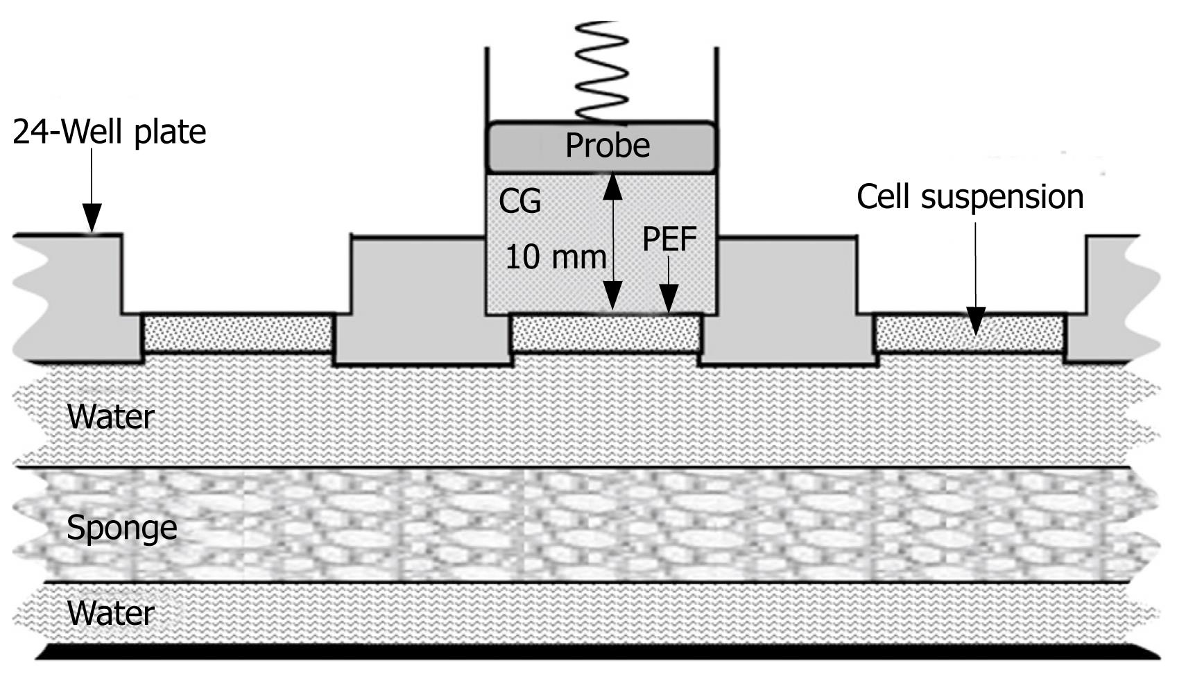
Figure 1 Schematic diagram of gene transfection.
Transducer with contact gel (CG) was placed on the cell suspension covered with a 6-&mgr;m poly-ester film (PEF) under 1.9 MHz continuous ultrasound with a 80.0 mW/cm2 output intensity. The 24-well plate was kept in a water bath at 37°C, and a sponge mat was placed in the water bath.
- Citation: Wang Y, Xu HX, Lu MD, Tang Q. Expression of thymidine kinase mediated by a novel non-viral delivery system under the control of vascular endothelial growth factor receptor 2 promoter selectively kills human umbilical vein endothelial cells. World J Gastroenterol 2008; 14(2): 224-230
- URL: https://www.wjgnet.com/1007-9327/full/v14/i2/224.htm
- DOI: https://dx.doi.org/10.3748/wjg.14.224


