Copyright
©2008 The WJG Press and Baishideng.
World J Gastroenterol. Jan 14, 2008; 14(2): 218-223
Published online Jan 14, 2008. doi: 10.3748/wjg.14.218
Published online Jan 14, 2008. doi: 10.3748/wjg.14.218
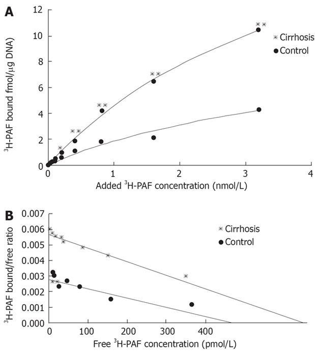
Figure 3 A: Saturation curve of 3H-PAF binding to cultured cirrhotic hepatic stellate cells.
3H-PAF in the concentration between 0.125 and 3.2 nmol/L,in presence or the absence of 5 &mgr;mol/L unlabeled incubated at 25°C for 3 h; B: Scatchard plot analysis of binding of 3H-PAF to cirrhotic hepatic stellate cells. Cirrhosis: Kd: 4.66 nmol/L, Bax: 24.65 ± 1.96 fmol/&mgr;g DNA, R = 0.982; Control: Kd: 3.51 nmol/L, Bax: 5.74 ± 1.55 fmol/&mgr;g DNA, R = 0.93.
- Citation: Chen Y, Wang CP, Lu YY, Zhou L, Su SH, Jia HJ, Feng YY, Yang YP. Hepatic stellate cells may be potential effectors of platelet activating factor induced portal hypertension. World J Gastroenterol 2008; 14(2): 218-223
- URL: https://www.wjgnet.com/1007-9327/full/v14/i2/218.htm
- DOI: https://dx.doi.org/10.3748/wjg.14.218


