Copyright
©2008 The WJG Press and Baishideng.
World J Gastroenterol. May 14, 2008; 14(18): 2924-2927
Published online May 14, 2008. doi: 10.3748/wjg.14.2924
Published online May 14, 2008. doi: 10.3748/wjg.14.2924
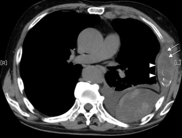
Figure 2 Chest CCT depicted a mass with necrosis inside the tumor (arrows) and enhancement of its border (arrowheads), and extrapleural tumor and pleural effusion.
- Citation: Matsuyama S, Shimonishi T, Yoshimura H, Higaki K, Nasu K, Toyooka M, Aoki S, Watanabe K, Sugihara H. An autopsy case of granulocyte-colony-stimulating-factor-producing extrahepatic bile duct carcinoma. World J Gastroenterol 2008; 14(18): 2924-2927
- URL: https://www.wjgnet.com/1007-9327/full/v14/i18/2924.htm
- DOI: https://dx.doi.org/10.3748/wjg.14.2924


