Copyright
©2008 The WJG Press and Baishideng.
World J Gastroenterol. May 14, 2008; 14(18): 2894-2899
Published online May 14, 2008. doi: 10.3748/wjg.14.2894
Published online May 14, 2008. doi: 10.3748/wjg.14.2894
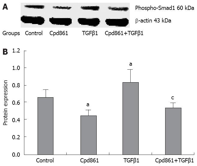
Figure 4 The level of Phosphorylated Smad1 with different treatment after 24 h.
Western blot was used as described. A: Representative Western blot results of Phosphorylated Smad1. The positions of protein size markers were given; B: Densitometry of Western-blot analyzed by Gel-pro software. The levels of Phospho-Smad1 were normalized to the level of β-actin protein. Six independent experiments were performed. aP < 0.05 vs untreated LX-2 cell group, cP < 0.05 vs TGFβ1 treated group. Error bars, SD.
- Citation: Li L, Zhao XY, Wang BE. Down-regulation of transforming growth factor β1/activin receptor-like kinase 1 pathway gene expression by herbal compound 861 is related to deactivation of LX-2 cells. World J Gastroenterol 2008; 14(18): 2894-2899
- URL: https://www.wjgnet.com/1007-9327/full/v14/i18/2894.htm
- DOI: https://dx.doi.org/10.3748/wjg.14.2894


