Copyright
©2008 The WJG Press and Baishideng.
World J Gastroenterol. May 14, 2008; 14(18): 2882-2887
Published online May 14, 2008. doi: 10.3748/wjg.14.2882
Published online May 14, 2008. doi: 10.3748/wjg.14.2882
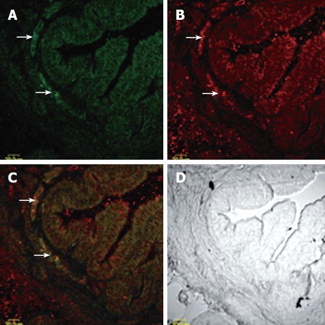
Figure 1 Overview of CCKAR-immunoreactive ICC seen on cross section of guinea pig gallbladder.
A: Low-magnification montage showing C-kit positive cells (arrow) distributed over a band of gallbladder muscle adjacent to the mucosa; B: Low-power montage showing CCKAR-immunoreactive cells distribute mainly in the muscular layers of guinea pig gallbladder; C: Compound micrographs from (A) and (B). Some, but not all C-kit immunoreactive (green) ICC colocalize CCKAR (red), meanwhile, not all CCKAR-immunoreactive cells are ICC; D: Phase-contrast micrographs of the same gallbladders wall at the same magnification.
- Citation: Xu D, Yu BP, Luo HS, Chen LD. Control of gallbladder contractions by cholecystokinin through cholecystokinin-A receptors on gallbladder interstitial cells of cajal. World J Gastroenterol 2008; 14(18): 2882-2887
- URL: https://www.wjgnet.com/1007-9327/full/v14/i18/2882.htm
- DOI: https://dx.doi.org/10.3748/wjg.14.2882


