Copyright
©2008 The WJG Press and Baishideng.
World J Gastroenterol. May 7, 2008; 14(17): 2748-2756
Published online May 7, 2008. doi: 10.3748/wjg.14.2748
Published online May 7, 2008. doi: 10.3748/wjg.14.2748
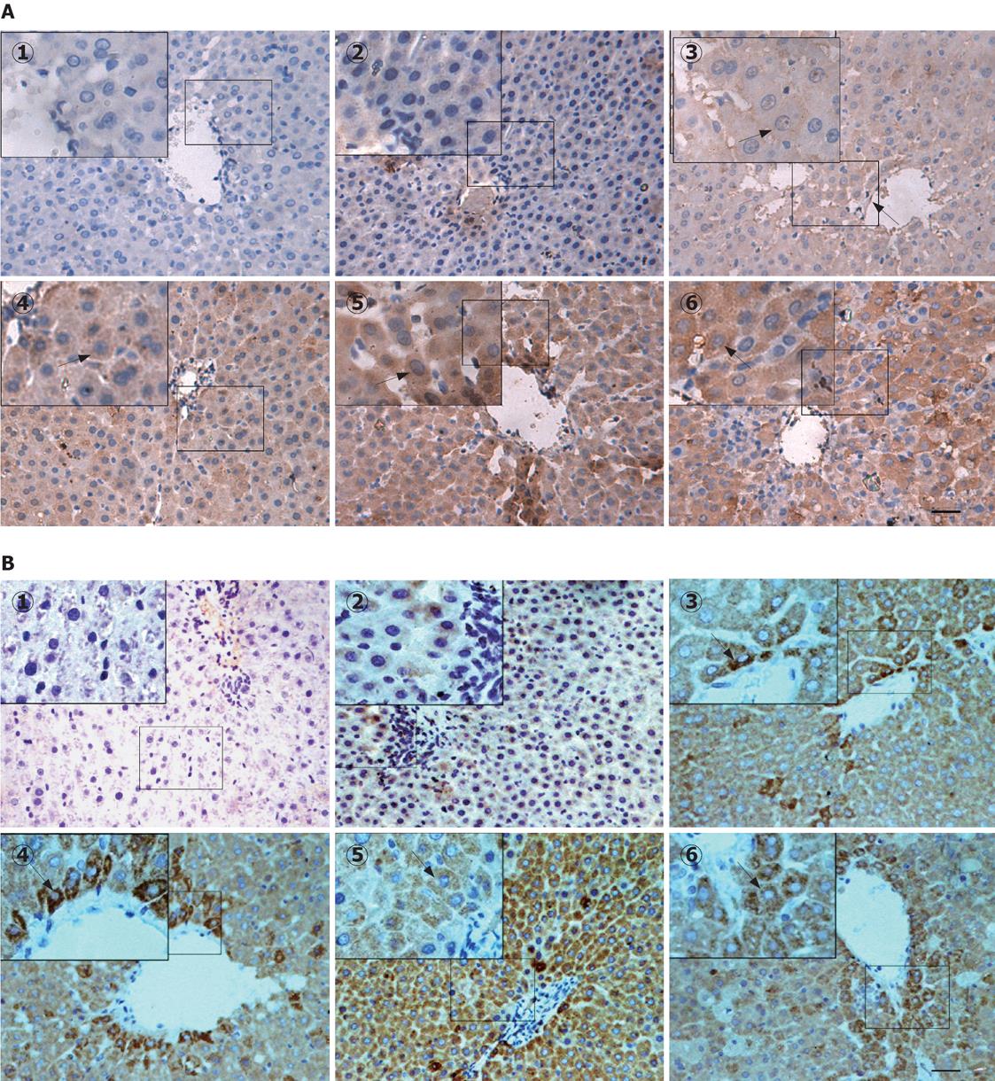
Figure 4 Immunohistochemical assay of the expression and localization of LTC4S (A) and mGST2 (B) in control and D-GalN/LPS-treated liver tissues.
Immunohistochemical staining for paraffin-embedded liver sections from the control and D-GalN/LPS-treated rats was examined as described in Materials and Methods. Arrows indicate the representative positive cells expressing the brown granules. 1: Absence of staining on omission of primary antibody; 2: Control; 3-6: Groups treated with D-GalN/LPS at 1, 3, 6 and 12 h, respectively. Bar = 100 &mgr;m.
- Citation: Ma KF, Yang HY, Chen Z, Qi LY, Zhu DY, Lou YJ. Enhanced expressions and activations of leukotriene C4 synthesis enzymes in D-galactosamine/lipopolysaccharide-induced rat fulminant hepatic failure model. World J Gastroenterol 2008; 14(17): 2748-2756
- URL: https://www.wjgnet.com/1007-9327/full/v14/i17/2748.htm
- DOI: https://dx.doi.org/10.3748/wjg.14.2748


