Copyright
©2008 The WJG Press and Baishideng.
World J Gastroenterol. Apr 28, 2008; 14(16): 2593-2595
Published online Apr 28, 2008. doi: 10.3748/wjg.14.2593
Published online Apr 28, 2008. doi: 10.3748/wjg.14.2593
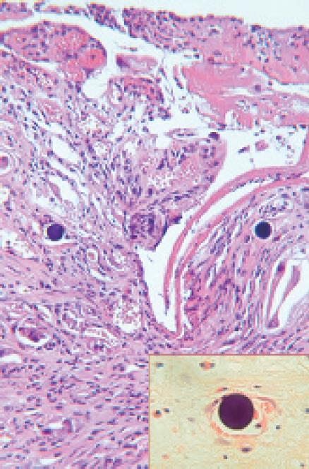
Figure 2 Gastric biopsy showing ulceration and typical inflammatory eschar at the surface.
The mucosa contains granulation tissue with inflammatory cells and several small blood vessels. Two round microspheres can be seen on the right and left side of the figure. Inset: 90Y-microsphere within a capillary adjacent to an erythrocyte.
- Citation: Mallach S, Ramp U, Erhardt A, Schmitt M, Häussinger D. An uncommon cause of gastro-duodenal ulceration. World J Gastroenterol 2008; 14(16): 2593-2595
- URL: https://www.wjgnet.com/1007-9327/full/v14/i16/2593.htm
- DOI: https://dx.doi.org/10.3748/wjg.14.2593


