Copyright
©2008 The WJG Press and Baishideng.
World J Gastroenterol. Apr 28, 2008; 14(16): 2593-2595
Published online Apr 28, 2008. doi: 10.3748/wjg.14.2593
Published online Apr 28, 2008. doi: 10.3748/wjg.14.2593
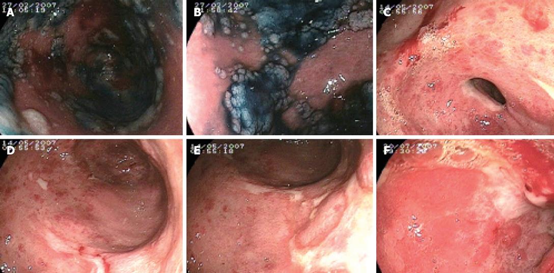
Figure 1 Chromoendoscopy.
A-B: Chromoendoscopy with 0.6% Kongored stain showing initial mucosal damage of the gastric antrum; C-E: Multiple ulcerations (Forrest III) and severe mucosal inflammation of the gastric antrum, 3 mo after SIRT; F: Partially reepithelialized ulcer (Forrest III) in the duodenal bulb, 5 mo after SIRT.
- Citation: Mallach S, Ramp U, Erhardt A, Schmitt M, Häussinger D. An uncommon cause of gastro-duodenal ulceration. World J Gastroenterol 2008; 14(16): 2593-2595
- URL: https://www.wjgnet.com/1007-9327/full/v14/i16/2593.htm
- DOI: https://dx.doi.org/10.3748/wjg.14.2593


