Copyright
©2008 The WJG Press and Baishideng.
World J Gastroenterol. Apr 14, 2008; 14(14): 2270-2271
Published online Apr 14, 2008. doi: 10.3748/wjg.14.2270
Published online Apr 14, 2008. doi: 10.3748/wjg.14.2270
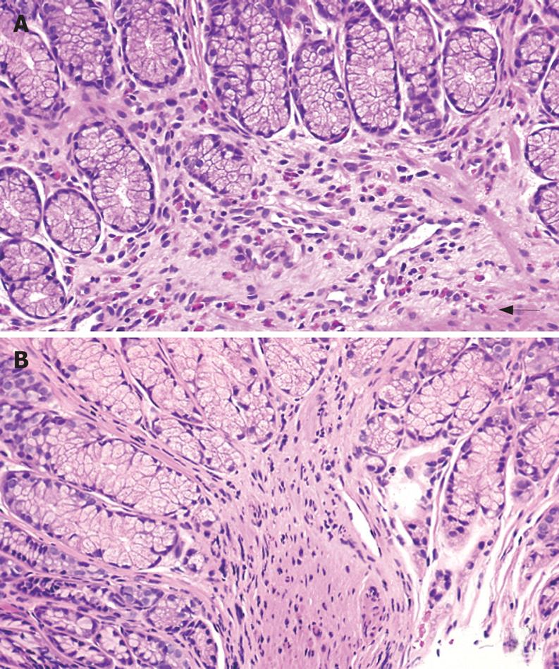
Figure 2 Histologic images before and 5 d after steroid therapy.
A: Peripyloric antral sections showed prominent eosinophilic infiltration of the lamina propria (up to 30 eosinophils per single high power field), with occasional degranulation (arrow) of eosinophilic content and infiltration of the muscularis mucosae; B: Biopsies 5 d after intravenous steroid therapy demonstrated only a few eosinophils with a peak count of 2 eosinophils per high power field (HE, × 40).
- Citation: Kellermayer R, Tatevian N, Klish W, Shulman RJ. Steroid responsive eosinophilic gastric outlet obstruction in a child. World J Gastroenterol 2008; 14(14): 2270-2271
- URL: https://www.wjgnet.com/1007-9327/full/v14/i14/2270.htm
- DOI: https://dx.doi.org/10.3748/wjg.14.2270


