Copyright
©2008 The WJG Press and Baishideng.
World J Gastroenterol. Apr 14, 2008; 14(14): 2208-2212
Published online Apr 14, 2008. doi: 10.3748/wjg.14.2208
Published online Apr 14, 2008. doi: 10.3748/wjg.14.2208
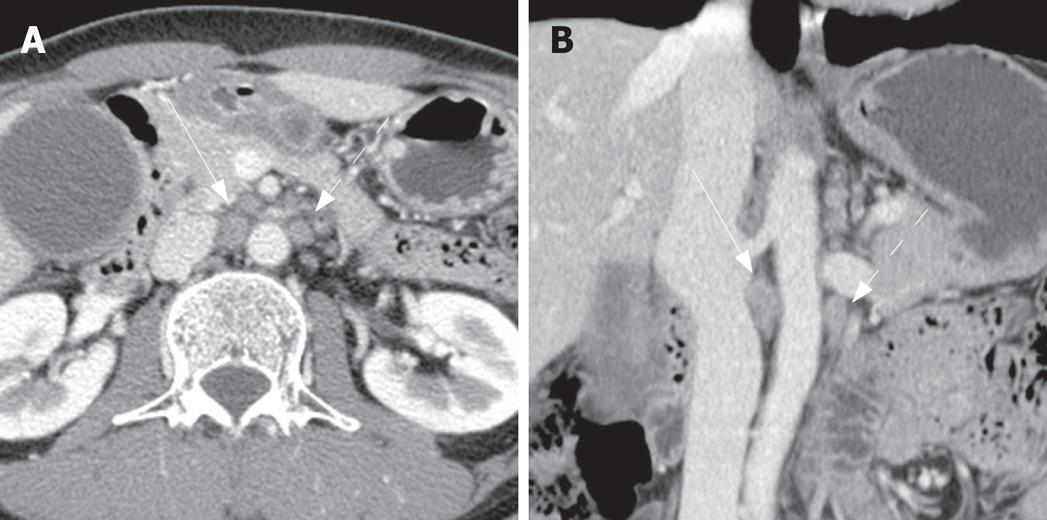
Figure 3 Two metastatic paraaortic lymph nodes in a 49-year-old man with gallbladder cancer.
Axial (A) and coronal (B) contrast-enhanced CT shows several paraaortic lymph nodes. Among them, the right largest node (straight arrow) shows 10 mm and 18.8 mm of short and long axis diameters with irregular margin (on coronal image), compatible with metastatic node. The left one (dot arrow) shows 8.2 mm and 12.2 mm of short and long axis diameters, less than the mean value of metastatic ones (9.2 mm and 13.2 mm, respectively). According to the best cut-off value of short diameter more than 5.3 mm and long axis diameter more than 11.6 mm, The left one is also metastatic one rather than non-metastatic one. Pathologic examination revealed that two lymph nodes were metastatic ones among six resected paraaortic lymph nodes.
- Citation: Kim YC, Park MS, Cha SW, Chung YE, Lim JS, Kim KS, Kim MJ, Kim KW. Comparison of CT and MRI for presurgical characterization of paraaortic lymph nodes in patients with pancreatico-biliary carcinoma. World J Gastroenterol 2008; 14(14): 2208-2212
- URL: https://www.wjgnet.com/1007-9327/full/v14/i14/2208.htm
- DOI: https://dx.doi.org/10.3748/wjg.14.2208


