Copyright
©2008 The WJG Press and Baishideng.
World J Gastroenterol. Apr 14, 2008; 14(14): 2194-2199
Published online Apr 14, 2008. doi: 10.3748/wjg.14.2194
Published online Apr 14, 2008. doi: 10.3748/wjg.14.2194
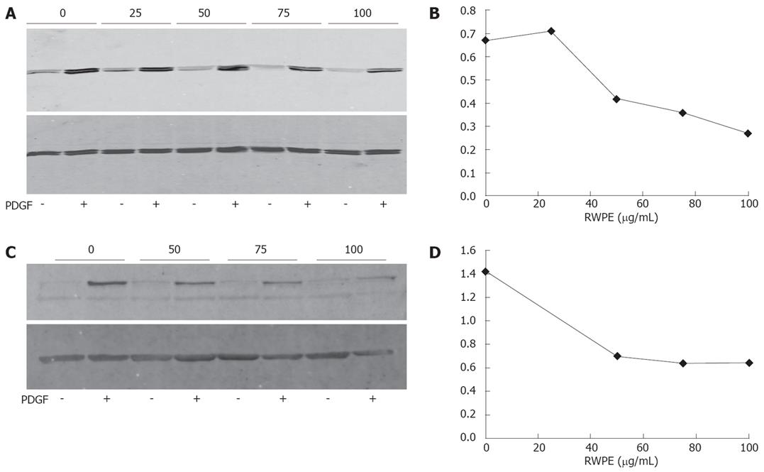
Figure 3 Effect of the RWPE on the phosphorylation of MAPK and Akt.
A: Myofibroblasts were pre-incubated for 1 h with the indicated concentrations of RWPE (in &mgr;g/mL) or solvent, then exposed for 10 min to 20 ng/mL PDGF-BB or buffer. Identical amounts of cell extracts were analyzed by Western blot with antibodies to phospho-ERK1/ERK2 (top panel) and to total ERKs (bottom panel). The picture is representative of 3 experiments; B: Quantitative analysis of the experiment shown in (A). The activation index refers to the ratio between the levels of phospho-ERK to those of total ERK; C: Same as in A except that the blot was labelled with an anti-phospho-Akt antibody (top panel) and an antibody to β-actin (bottom panel); D: Quantitative analysis of the experiment shown in (C). The activation index refers to the ratio between the levels of phospho-Akt to those of β-actin.
- Citation: Neaud V, Rosenbaum J. A red wine polyphenolic extract reduces the activation phenotype of cultured human liver myofibroblasts. World J Gastroenterol 2008; 14(14): 2194-2199
- URL: https://www.wjgnet.com/1007-9327/full/v14/i14/2194.htm
- DOI: https://dx.doi.org/10.3748/wjg.14.2194


