Copyright
©2008 The WJG Press and Baishideng.
World J Gastroenterol. Apr 7, 2008; 14(13): 2085-2088
Published online Apr 7, 2008. doi: 10.3748/wjg.14.2085
Published online Apr 7, 2008. doi: 10.3748/wjg.14.2085
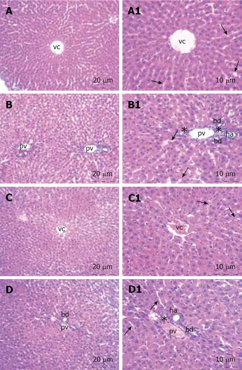
Figure 1 Liver sections stained with hematoxyline and eosin.
A, B: Control group showing the regular architecture; C, D: Treatment group. vc: Central vein; pv: Portal vein; ha: Hepatic artery; bd: Bile ductules. Arrow, hepatic sinusoids; *, cell infiltration. The right column of the panel is the larger view of the left column.
- Citation: Kilicoglu B, Kismet K, Kilicoglu SS, Erel S, Gencay O, Sorkun K, Erdemli E, Akhan O, Akkus MA, Sayek I. Effects of honey as a scolicidal agent on the hepatobiliary system. World J Gastroenterol 2008; 14(13): 2085-2088
- URL: https://www.wjgnet.com/1007-9327/full/v14/i13/2085.htm
- DOI: https://dx.doi.org/10.3748/wjg.14.2085


