Copyright
©2008 The WJG Press and Baishideng.
World J Gastroenterol. Mar 28, 2008; 14(12): 1858-1865
Published online Mar 28, 2008. doi: 10.3748/wjg.14.1858
Published online Mar 28, 2008. doi: 10.3748/wjg.14.1858
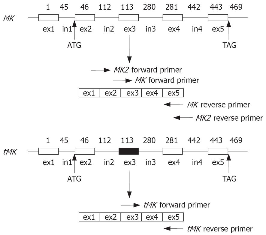
Figure 1 Illustration of MK and tMK gene DNA structures.
Box: Exon (ex); Line: Intron (in); Shaded box; Truncated portion; ATG: Start site; TAG: Terminal site; Numeric figures: Nucleotide position of the mRNA transcript. Arrowheads indicate the sites of primer complemented with MK or tMK mRNA.
-
Citation: Wang QL, Wang H, Zhao SL, Huang YH, Hou YY. Over-expressed and truncated midkines promote proliferation of BGC823 cells
in vitro and tumor growthin vivo . World J Gastroenterol 2008; 14(12): 1858-1865 - URL: https://www.wjgnet.com/1007-9327/full/v14/i12/1858.htm
- DOI: https://dx.doi.org/10.3748/wjg.14.1858


