Copyright
©2008 The WJG Press and Baishideng.
World J Gastroenterol. Mar 21, 2008; 14(11): 1768-1773
Published online Mar 21, 2008. doi: 10.3748/wjg.14.1768
Published online Mar 21, 2008. doi: 10.3748/wjg.14.1768
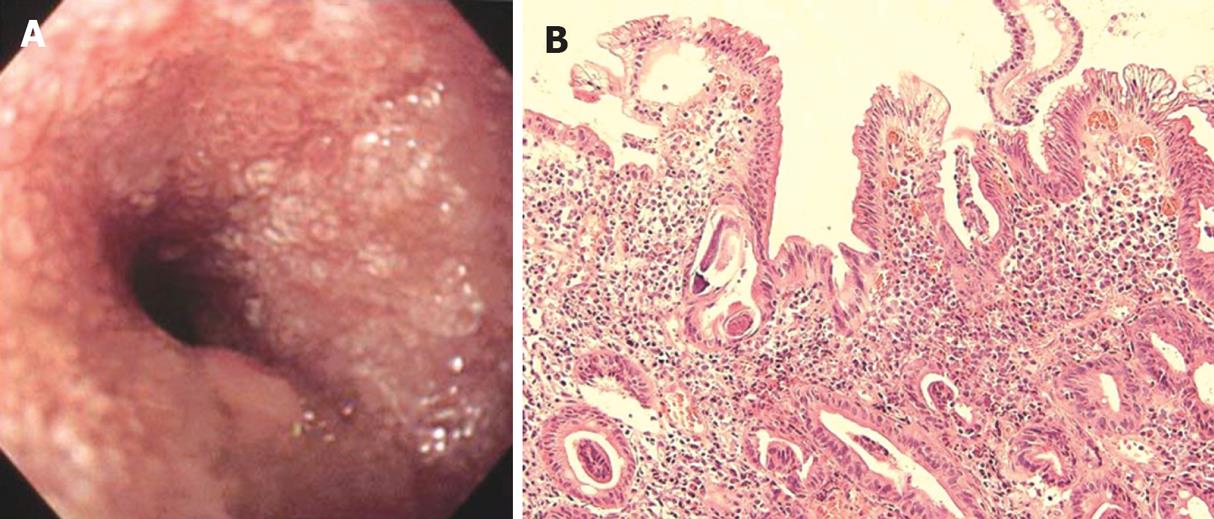
Figure 4 Endoscopic image and HE staining of duodenal biopsy.
A: EGD showing white villi and stenosis in the second part of duodenal (Case 16); B: Biopsy specimen from the mucosa showing numerous larvae with severe villous atrophy and moderate inflammatory cell infiltration (HE, × 200).
-
Citation: Kishimoto K, Hokama A, Hirata T, Ihama Y, Nakamoto M, Kinjo N, Kinjo F, Fujita J. Endoscopic and histopathological study on the duodenum of
Strongyloides stercoralis hyperinfection. World J Gastroenterol 2008; 14(11): 1768-1773 - URL: https://www.wjgnet.com/1007-9327/full/v14/i11/1768.htm
- DOI: https://dx.doi.org/10.3748/wjg.14.1768


