Copyright
©2008 The WJG Press and Baishideng.
World J Gastroenterol. Mar 21, 2008; 14(11): 1759-1767
Published online Mar 21, 2008. doi: 10.3748/wjg.14.1759
Published online Mar 21, 2008. doi: 10.3748/wjg.14.1759
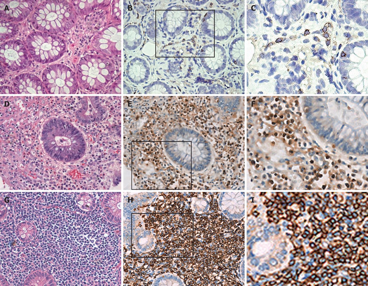
Figure 2 Immunohistochemical analysis of the expression of NFAT2 in colon mucosa tissues.
A-C are derived from a case of normal control, D-F from a case of UC, and G-I from a case of CD. A, D ,G are the results of H&E stain in 100 × magnification. B, E, H are the results of immunohistochemistry for NFAT2 counter-staining with hematoxylin for nuclei, 100 × magnification. C, F, I are the close view of B, E, H respectively. Of note, NFAT2 was exclusively located in cytoplasm of LMPCs of normal colon mucosa as well as LMPCs of CD affected colonic mucosa, whereas NFAT2 was primarily located in the nuclei of LMPCs of the UC affected colonic mucosa.
- Citation: Shih TC, Hsieh SY, Hsieh YY, Chen TC, Yeh CY, Lin CJ, Lin DY, Chiu CT. Aberrant activation of nuclear factor of activated T cell 2 in lamina propria mononuclear cells in ulcerative colitis. World J Gastroenterol 2008; 14(11): 1759-1767
- URL: https://www.wjgnet.com/1007-9327/full/v14/i11/1759.htm
- DOI: https://dx.doi.org/10.3748/wjg.14.1759


