Copyright
©2008 The WJG Press and Baishideng.
World J Gastroenterol. Mar 14, 2008; 14(10): 1625-1629
Published online Mar 14, 2008. doi: 10.3748/wjg.14.1625
Published online Mar 14, 2008. doi: 10.3748/wjg.14.1625
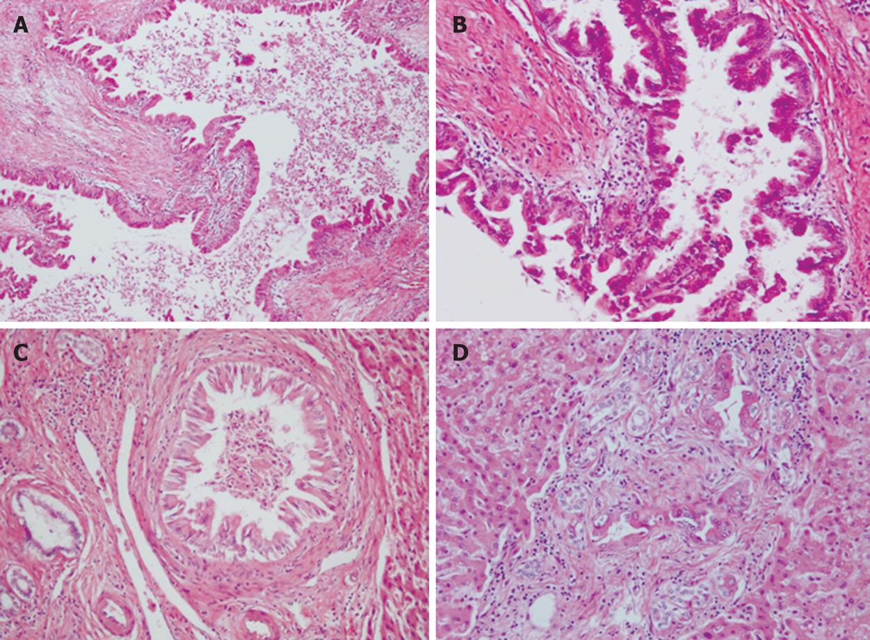
Figure 5 Histopathological findings showing.
A: A view of the intrahepatic bile ducts dilated with peri-ductal fibrosis (× 100); B: Carcinoma cells continuously proliferating from the intrahepatic large bile ducts to small bile ducts (× 400); C: Carcinoma cells continuously proliferating from the intrahepatic large bile ducts to small bile ducts (× 200); D: Carcinoma cells in bile ductules around the small portal area (× 200).
- Citation: Hayashi J, Matsuoka SI, Inami M, Ohshiro S, Ishigami A, Fujikawa H, Miyagawa M, Mimatsu K, Kuboi Y, Kanou H, Oida T, Moriyama M. A case of asymptomatic intraductal papillary neoplasm of the bile duct without hepatolithiasis. World J Gastroenterol 2008; 14(10): 1625-1629
- URL: https://www.wjgnet.com/1007-9327/full/v14/i10/1625.htm
- DOI: https://dx.doi.org/10.3748/wjg.14.1625


