Copyright
©2008 The WJG Press and Baishideng.
World J Gastroenterol. Mar 14, 2008; 14(10): 1528-1533
Published online Mar 14, 2008. doi: 10.3748/wjg.14.1528
Published online Mar 14, 2008. doi: 10.3748/wjg.14.1528
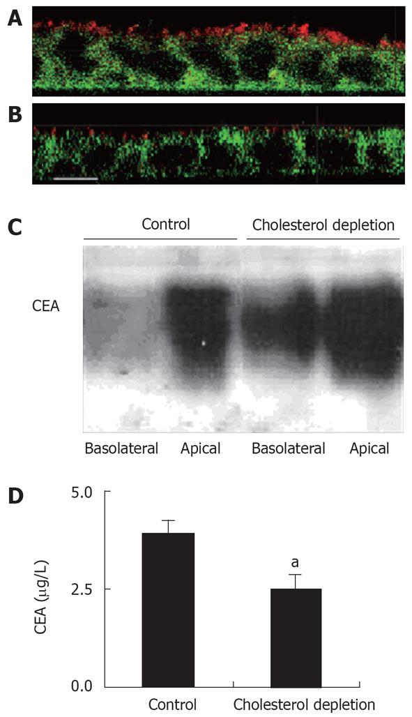
Figure 3 The effect of cholesterol depletion on apical and basolateral secretion of CEA.
A, B: X-Z confocal views of cells labeled with antibodies directed against CEA (red) and Na+/K+-ATPase (green). In non-depleted cells under steady state conditions, CEA family proteins are expressed at the apical surface (A). Caco-2 cells were grown to confluency on polycarbonate filters for 14 d and cholesterol-depleted with a combination of lovastatin/MβCD. After cholesterol depletion, CEA was still apical but with a lower expression level (B) but no basolateral staining of CEA was detected (Bar 10 &mgr;m). C: Media from apical and basolateral chamber were collected for 2 h and Western blots performed. In non-depleted cells, most of the CEA is secreted into the apical medium. In cholesterol depleted cells, a significant amount was also found in the basolateral medium. D: Quantification of apical CEA secretion using an ECLISA from Roche. Six similar experiments were performed. An asterisk indicates significant differences (3.12 ± 0.62 vs 1.91 ± 0.81, aP < 0.05).
- Citation: Ehehalt R, Krautter M, Zorn M, Sparla R, Füllekrug J, Kulaksiz H, Stremmel W. Increased basolateral sorting of carcinoembryonic antigen in a polarized colon carcinoma cell line after cholesterol depletion-Implications for treatment of inflammatory bowel disease. World J Gastroenterol 2008; 14(10): 1528-1533
- URL: https://www.wjgnet.com/1007-9327/full/v14/i10/1528.htm
- DOI: https://dx.doi.org/10.3748/wjg.14.1528


