Copyright
©2007 Baishideng Publishing Group Co.
World J Gastroenterol. Feb 28, 2007; 13(8): 1275-1278
Published online Feb 28, 2007. doi: 10.3748/wjg.v13.i8.1275
Published online Feb 28, 2007. doi: 10.3748/wjg.v13.i8.1275
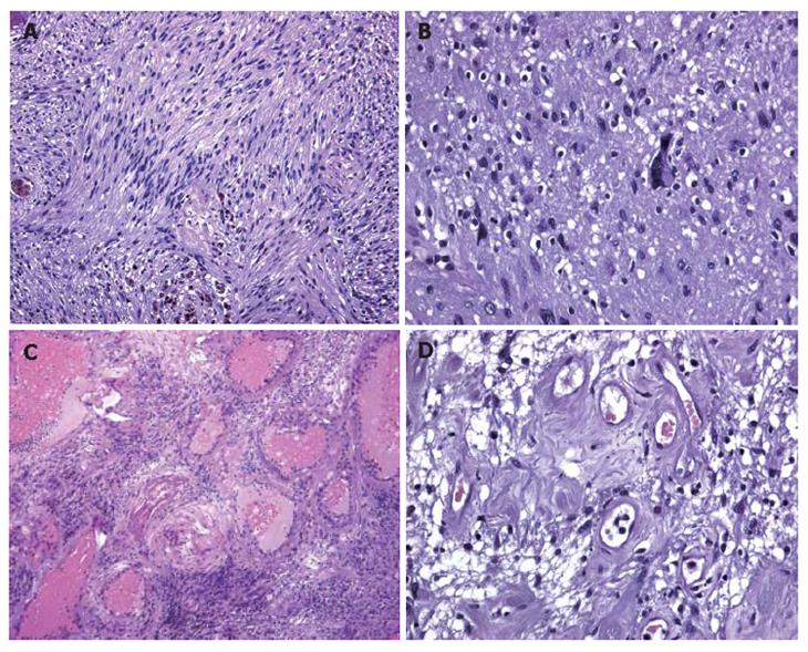
Figure 3 A: Histological examination showing that inter-woven bundles of fused cells separated by slacker, oedematous areas with a pseudocystic appearance (Antoni A schwannoma growth pattern); B: Histological examination showing a few cells with hyperchromic, atypical and bizarre nuclei.
Histological examination showing a few cells with hyperchromic, atypical and bizarre nuclei; C: Histological examination showing hyalinisation and ectatic and partially thrombosed vessels; D: Histological examination showing perivascular hyalinisation.
- Citation: Fenoglio L, Severini S, Cena P, Migliore E, Bracco C, Pomero F, Panzone S, Cavallero GB, Silvestri A, Brizio R, Borghi F. Common bile duct schwannoma: A case report and review of literature. World J Gastroenterol 2007; 13(8): 1275-1278
- URL: https://www.wjgnet.com/1007-9327/full/v13/i8/1275.htm
- DOI: https://dx.doi.org/10.3748/wjg.v13.i8.1275


