Copyright
©2007 Baishideng Publishing Group Co.
World J Gastroenterol. Feb 28, 2007; 13(8): 1271-1274
Published online Feb 28, 2007. doi: 10.3748/wjg.v13.i8.1271
Published online Feb 28, 2007. doi: 10.3748/wjg.v13.i8.1271
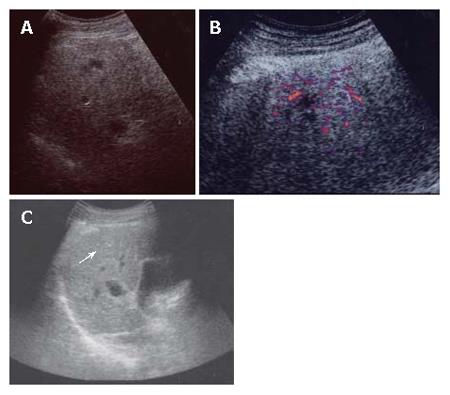
Figure 2 A: US imaging (April 2005).
A 10 mm hypoechoic nodule in S7; B: contrast enhanced US (April 2005). Hypovascularity in the early phase; C: US imaging (May 2006). A 10 mm hyperechoic nodule in S7.
- Citation: Kim SR, Ikawa H, Ando K, Mita K, Fuki S, Sakamoto M, Kanbara Y, Matsuoka T, Kudo M, Hayashi Y. Multistep hepatocarcinogenesis from a dysplastic nodule to well-differentiated hepatocellular carcinoma in a patient with alcohol-related liver cirrhosis. World J Gastroenterol 2007; 13(8): 1271-1274
- URL: https://www.wjgnet.com/1007-9327/full/v13/i8/1271.htm
- DOI: https://dx.doi.org/10.3748/wjg.v13.i8.1271


