Copyright
©2007 Baishideng Publishing Group Co.
World J Gastroenterol. Feb 14, 2007; 13(6): 960-963
Published online Feb 14, 2007. doi: 10.3748/wjg.v13.i6.960
Published online Feb 14, 2007. doi: 10.3748/wjg.v13.i6.960
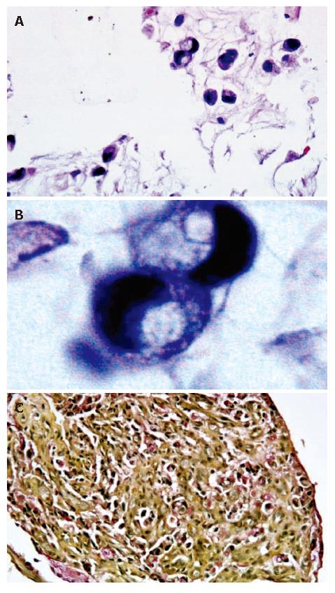
Figure 2 Histology and immunohistochemistry of biopsies from gastric: Light microscopy with hematoxylin and eosin staining of tumor tissue at 40X and 100X with oil magnifications demonstrating moderately-sized cells with abundant vacuolated cytoplasm (A) and occasional signet ring cells (B), and mucicarmine stain demonstrating intracytoplasmic positive staining (C).
- Citation: Ghosh P, Miyai K, Chojkier M. Gastric adenocarcinoma inducing portal hypertension: A rare presentation. World J Gastroenterol 2007; 13(6): 960-963
- URL: https://www.wjgnet.com/1007-9327/full/v13/i6/960.htm
- DOI: https://dx.doi.org/10.3748/wjg.v13.i6.960


