Copyright
©2007 Baishideng Publishing Group Co.
World J Gastroenterol. Feb 14, 2007; 13(6): 960-963
Published online Feb 14, 2007. doi: 10.3748/wjg.v13.i6.960
Published online Feb 14, 2007. doi: 10.3748/wjg.v13.i6.960
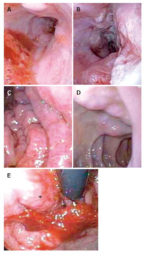
Figure 1 Esophagogastroduodenoscopy demonstratring grade 1 esophageal varices (A), tumor invasion of distal third of the esophagus (B), moderate portal hypertensive gastropathy (C), duodenal varices (D), retroflexed view of the nodular tumor in the gastric fundus (E).
- Citation: Ghosh P, Miyai K, Chojkier M. Gastric adenocarcinoma inducing portal hypertension: A rare presentation. World J Gastroenterol 2007; 13(6): 960-963
- URL: https://www.wjgnet.com/1007-9327/full/v13/i6/960.htm
- DOI: https://dx.doi.org/10.3748/wjg.v13.i6.960


