Copyright
©2007 Baishideng Publishing Group Co.
World J Gastroenterol. Feb 14, 2007; 13(6): 874-881
Published online Feb 14, 2007. doi: 10.3748/wjg.v13.i6.874
Published online Feb 14, 2007. doi: 10.3748/wjg.v13.i6.874
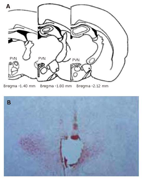
Figure 1 Sites of the stimulating electrode tip in PVN.
A: Standard atlas sections of the rat brain showing the distributions of stimulating sites of the experimental animals; B: Photomicrographs of stimulating sites in the rat brain. The section stained with neutral red, showing the position of stimulating electrode tip by passing a positive DC of 1 mA for 10 s, which indicates a placement within the PVN.
- Citation: Li L, Zhang YM, Qiao WL, Wang L, Zhang JF. Effects of hypothalamic paraventricular nuclei on apoptosis and proliferation of gastric mucosal cells induced by ischemia/reperfusion in rats. World J Gastroenterol 2007; 13(6): 874-881
- URL: https://www.wjgnet.com/1007-9327/full/v13/i6/874.htm
- DOI: https://dx.doi.org/10.3748/wjg.v13.i6.874


