Copyright
©2007 Baishideng Publishing Group Co.
World J Gastroenterol. Feb 14, 2007; 13(6): 858-865
Published online Feb 14, 2007. doi: 10.3748/wjg.v13.i6.858
Published online Feb 14, 2007. doi: 10.3748/wjg.v13.i6.858
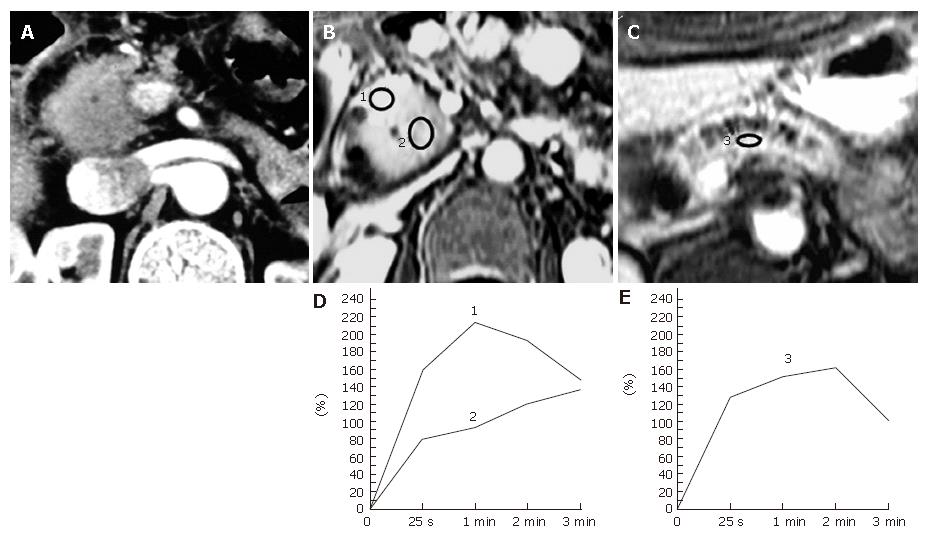
Figure 3 Pancreatic carcinoma occurring in a 67-year-old man with a longstanding chronic pancreatitis.
A: An abdominal contrast-enhanced CT image shows a focal enlargement of the head of the pancreas. The tumor-to-parenchymal attenuation difference is obscure. The patient underwent a laparotomy under a diagnosis of tumor-forming pancreatitis presenting with obstructive jaundice and was found to have pancreas head carcinoma during the operation; B, C: Dynamic contrast-enhanced MRI images of the pancreas. The ROIs are placed at the focally enlarged pancreas head (No.2 ROI), the proximal side of the head of the pancreas (No.1 ROI) , and the body of the pancreas (No.3 ROI); D: Pancreatic TICs obtained from the No.1 and No.2 ROIs as in (B) demonstrate type-II and type-IV, respectively; E: Pancreatic TIC obtained from the No.3 ROI as in (C) shows type-III.
- Citation: Tajima Y, Kuroki T, Tsutsumi R, Isomoto I, Uetani M, Kanematsu T. Pancreatic carcinoma coexisting with chronic pancreatitis versus tumor-forming pancreatitis: Diagnostic utility of the time-signal intensity curve from dynamic contrast-enhanced MR imaging. World J Gastroenterol 2007; 13(6): 858-865
- URL: https://www.wjgnet.com/1007-9327/full/v13/i6/858.htm
- DOI: https://dx.doi.org/10.3748/wjg.v13.i6.858


