Copyright
©2007 Baishideng Publishing Group Co.
World J Gastroenterol. Feb 14, 2007; 13(6): 821-825
Published online Feb 14, 2007. doi: 10.3748/wjg.v13.i6.821
Published online Feb 14, 2007. doi: 10.3748/wjg.v13.i6.821
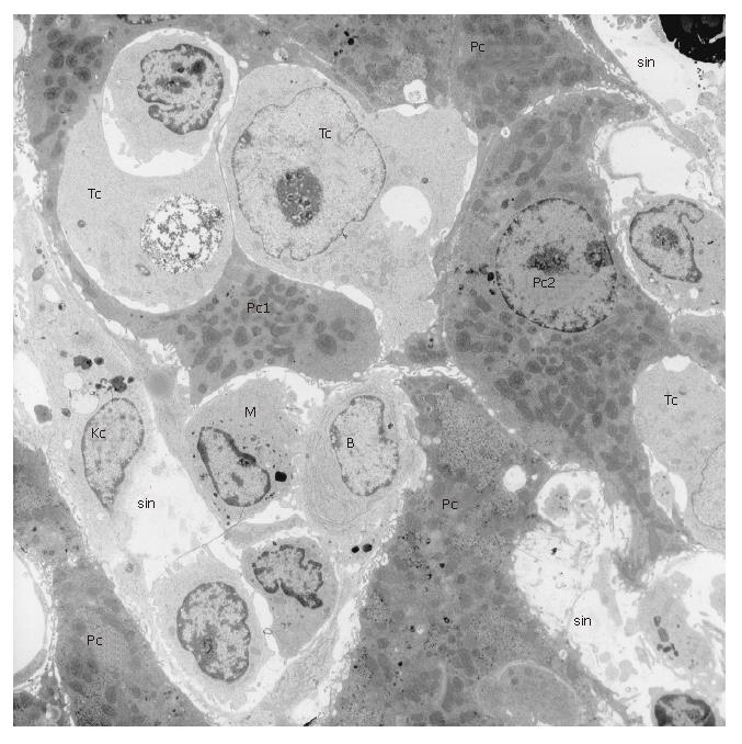
Figure 2 Transmission electron microscopy photomicrograph at low magnification (6700 x) of rat liver one week after i.
v. injection of one million CC531s colon cancer cells. Tumour cells (Tc) can be recognized by a ribosome-rich, bulky cytoplasm containing few organelles. Tumour cell nuclei are large and contain mostly euchromatin. In this picture, tumour cells have already taken position in the liver parenchyma and compress or deform the parenchymal cells (Pc1 and Pc2). In this stage, tumour cells are so numerous that defending cells, such as Kupffer cells (Kc) and monocytes (M) in the hepatic sinusoids (sin) and B lymphocytes (B) seem to be no longer involved as in earlier stages. At this stage of tumour cell settlement, rebuilding of the hepatic tissue sets in and soon small visible white spots of metastasis will be seen on the surface of the liver.
- Citation: Braet F, Nagatsuma K, Saito M, Soon L, Wisse E, Matsuura T. The hepatic sinusoidal endothelial lining and colorectal liver metastases. World J Gastroenterol 2007; 13(6): 821-825
- URL: https://www.wjgnet.com/1007-9327/full/v13/i6/821.htm
- DOI: https://dx.doi.org/10.3748/wjg.v13.i6.821


