Copyright
©2007 Baishideng Publishing Group Co.
World J Gastroenterol. Feb 7, 2007; 13(5): 683-691
Published online Feb 7, 2007. doi: 10.3748/wjg.v13.i5.683
Published online Feb 7, 2007. doi: 10.3748/wjg.v13.i5.683
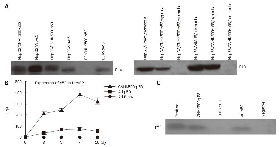
Figure 3 A: E1A and E1B expression identified by Western blot demonstrating that all HCC cells infected with CNHK500-p53 or WtAd5 were positive for E1A expression, however, normal BJ cells were negative for E1A expression when they were infected with CNHK500-p53, and positive only when they were infected with WtAd5, while E1B of CNHK500-p53 was only expressed under hypoxia condition in HepG2 and Hep3B, and expressed both under normal and hypoxia condition with WtAd5; B: ELISA assay showing that p53 protein secreted from a HepG2, infected with CNHK500-p53, was significantly higher than that infected with nonreplicative adenovirus Ad-p53 and Ad-Blank in vitro (P < 0.
05); C: Western blot showing enhanced p53 expression in HepG2 infected with CNHK500-p53.
-
Citation: Zhao HC, Zhang Q, Yang Y, Lu MQ, Li H, Xu C, Chen GH. p53-expressing conditionally replicative adenovirus CNHK500-p53 against hepatocellular carcinoma
in vitro . World J Gastroenterol 2007; 13(5): 683-691 - URL: https://www.wjgnet.com/1007-9327/full/v13/i5/683.htm
- DOI: https://dx.doi.org/10.3748/wjg.v13.i5.683


