Copyright
©2007 Baishideng Publishing Group Co.
World J Gastroenterol. Dec 28, 2007; 13(48): 6538-6548
Published online Dec 28, 2007. doi: 10.3748/wjg.v13.i48.6538
Published online Dec 28, 2007. doi: 10.3748/wjg.v13.i48.6538
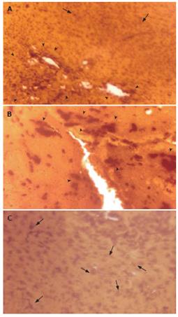
Figure 4 Light micrographs of tissue sections from murine liver (after 30 d of tumour transplantation) showing immunostaining of metallothionein (MT) with anti-rat MT-1 antibody and 3, 3'-diaminobenzidine tetrahydrochloride (DAB).
A and B: Strong immunostaining of MT protein in EAC Control mice; C: Reduced immunostaining of MT in EAC + ALE-treatment mice. ALE: Aqueous leaf extract of A. ilicifolius; EAC: Ehrlich ascites carcinoma. Arrow Head ( ) indicates intense immunostaining of MT protein with prominent focal expression and isolated clusters of MT-positive cells. Arrow (↑) indicates scattered/individual MT immunopositive cells. Magnification, A: × 100; B and C: × 270.
-
Citation: Chakraborty T, Bhuniya D, Chatterjee M, Rahaman M, Singha D, Chatterjee BN, Datta S, Rana A, Samanta K, Srivastawa S, Maitra SK, Chatterjee M.
Acanthus ilicifolius plant extract prevents DNA alterations in a transplantable Ehrlich ascites carcinoma-bearing murine model. World J Gastroenterol 2007; 13(48): 6538-6548 - URL: https://www.wjgnet.com/1007-9327/full/v13/i48/6538.htm
- DOI: https://dx.doi.org/10.3748/wjg.v13.i48.6538


