Copyright
©2007 Baishideng Publishing Group Co.
World J Gastroenterol. Dec 28, 2007; 13(48): 6538-6548
Published online Dec 28, 2007. doi: 10.3748/wjg.v13.i48.6538
Published online Dec 28, 2007. doi: 10.3748/wjg.v13.i48.6538
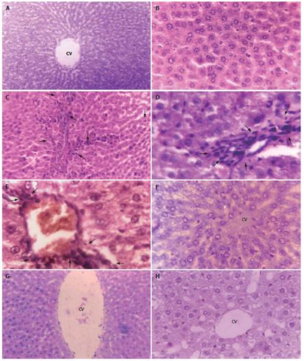
Figure 3 Contiguous liver sections from mice showing hepatocellular histological profiles.
A, B, F: Normal hepatocellular architecture depicting hepatocytes radiating from the central vein; C, D, E: Aberrant hepatocellular phenotype with prominent basophilic focal lesions (black arrows) and the presence of eosinophilic and clear cell foci following serial, intraperitoneal (ip) inoculations of viable Ehrlich ascites carcinoma (EAC) cells; G, H: Almost normal hepatocellular architecture following simultaneous ip administrations of aqueous leaf extract (ALE) of A. ilicifolius and EAC. A and B: Untreated Control; C, D, and E: EAC Control; F: ALE Control; G and H: EAC + ALE. CV: Central vein. Magnification, A, C, and G: × 100; B, D, E, F, H: × 450.
-
Citation: Chakraborty T, Bhuniya D, Chatterjee M, Rahaman M, Singha D, Chatterjee BN, Datta S, Rana A, Samanta K, Srivastawa S, Maitra SK, Chatterjee M.
Acanthus ilicifolius plant extract prevents DNA alterations in a transplantable Ehrlich ascites carcinoma-bearing murine model. World J Gastroenterol 2007; 13(48): 6538-6548 - URL: https://www.wjgnet.com/1007-9327/full/v13/i48/6538.htm
- DOI: https://dx.doi.org/10.3748/wjg.v13.i48.6538


