Copyright
©2007 Baishideng Publishing Group Inc.
World J Gastroenterol. Dec 21, 2007; 13(47): 6436-6438
Published online Dec 21, 2007. doi: 10.3748/wjg.v13.i47.6436
Published online Dec 21, 2007. doi: 10.3748/wjg.v13.i47.6436
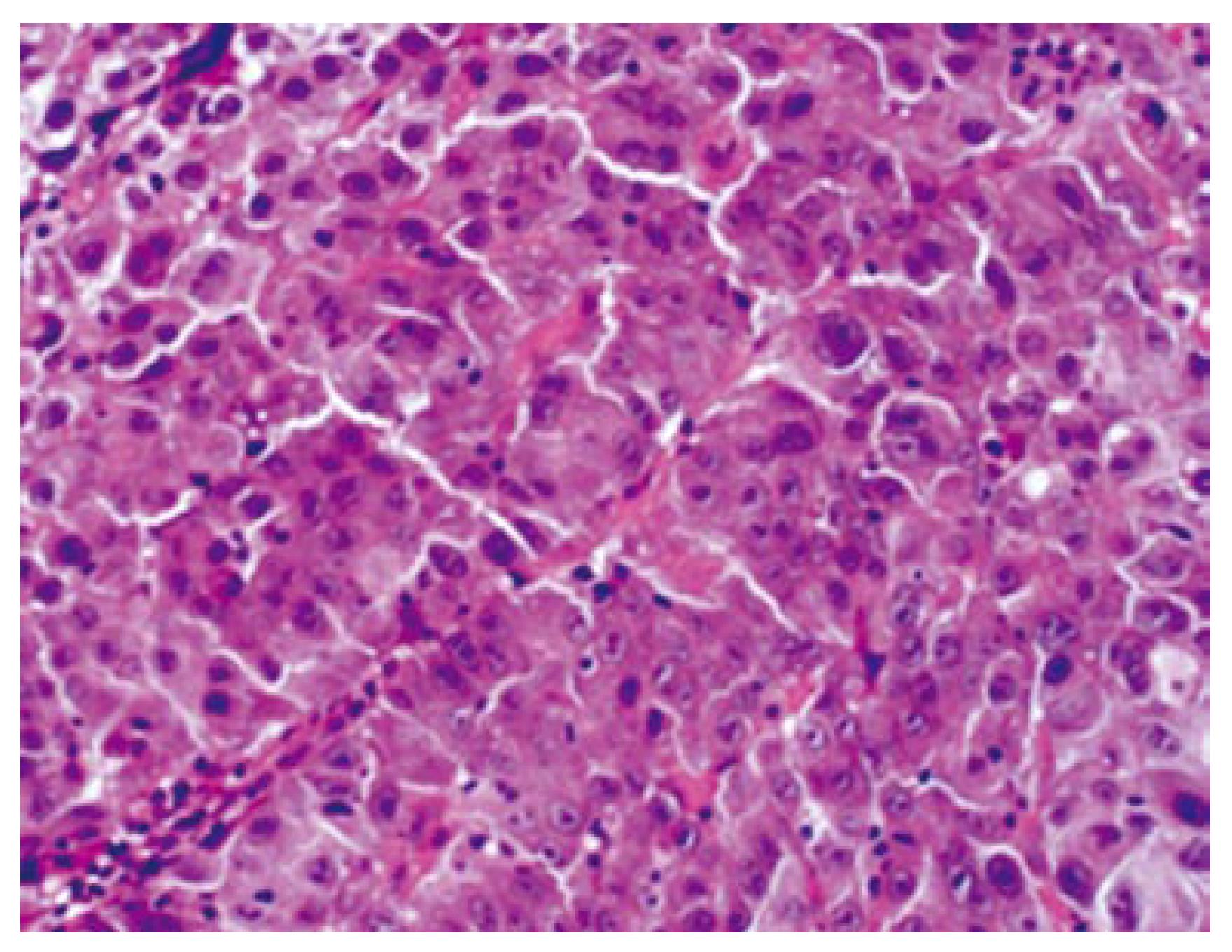
Figure 2 Microscopically, the lesion in the external auditory canal is composed of proliferating atypical epithelial cells forming a solid and thick trabecular arrangement with necrosis, and a fibrous stroma (hematoxylin/eosin stain, x 400).
- Citation: Yasumatsu R, Okura K, Sakiyama Y, Nakamuta M, Matsumura T, Uehara S, Yamamoto T, Komune S. Metastatic hepatocellular carcinoma of the external auditory canal. World J Gastroenterol 2007; 13(47): 6436-6438
- URL: https://www.wjgnet.com/1007-9327/full/v13/i47/6436.htm
- DOI: https://dx.doi.org/10.3748/wjg.v13.i47.6436


