Copyright
©2007 Baishideng Publishing Group Inc.
World J Gastroenterol. Dec 21, 2007; 13(47): 6404-6409
Published online Dec 21, 2007. doi: 10.3748/wjg.v13.i47.6404
Published online Dec 21, 2007. doi: 10.3748/wjg.v13.i47.6404
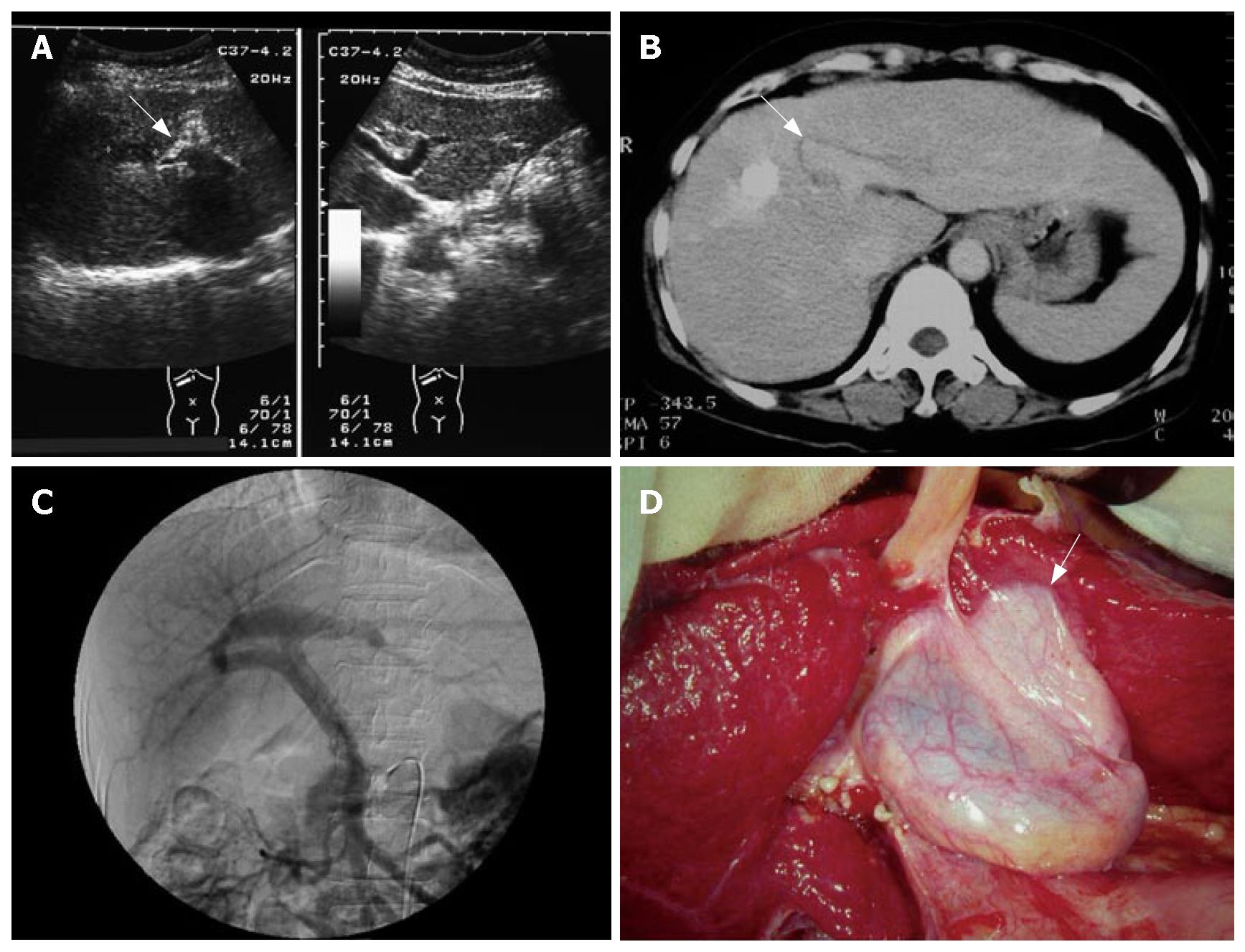
Figure 1 A 50-year-old woman with chronic hepatitis C.
Right trise-gmentectomy and cholecy-stectomy were performed due to the right lobe liver tumor. A: US demonstrated a club-shaped portal vein (arrow) over the right lobe of the liver and no umbilical portion over the left lobe of the liver; B: Enhanced CT demonstrated enlarged left lateral segment and a club-shaped portal vein (arrow) over the right side of the liver; C: Angiography demonstrated trifurcation type of the portal vein; D: Intraoperative photography demo-nstrated gallbladder over left side of ligamentum teres and adhesions on the left lateral segment (arrow).
- Citation: Hsu SL, Chen TY, Huang TL, Sun CK, Concejero AM, Tsang LLC, Cheng YF. Left-sided gallbladder: Its clinical significance and imaging presentations. World J Gastroenterol 2007; 13(47): 6404-6409
- URL: https://www.wjgnet.com/1007-9327/full/v13/i47/6404.htm
- DOI: https://dx.doi.org/10.3748/wjg.v13.i47.6404


