Copyright
©2007 Baishideng Publishing Group Inc.
World J Gastroenterol. Nov 21, 2007; 13(43): 5787-5793
Published online Nov 21, 2007. doi: 10.3748/wjg.v13.i43.5787
Published online Nov 21, 2007. doi: 10.3748/wjg.v13.i43.5787
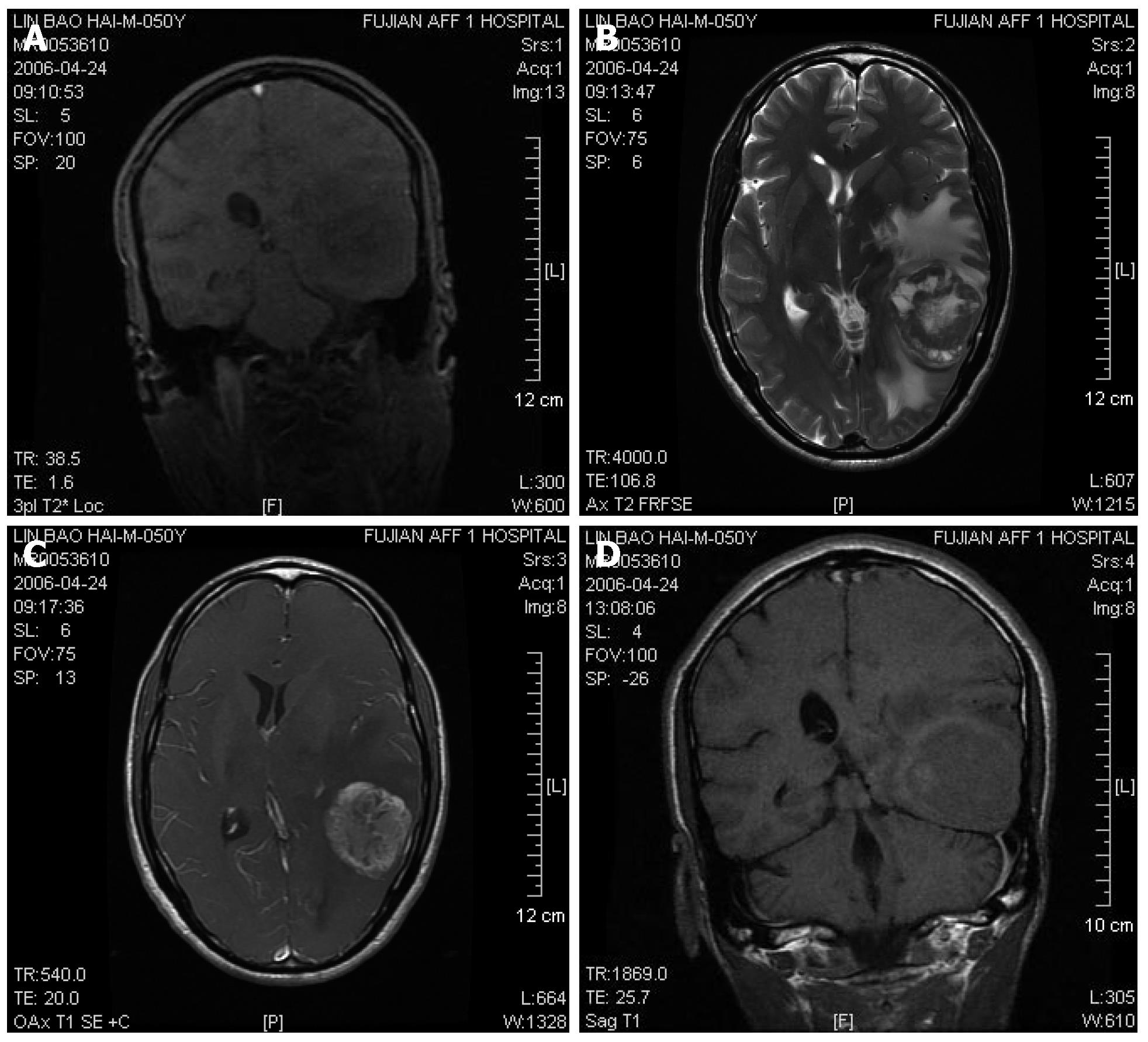
Figure 1 MR imaging revealing a spherical occupying lesion in the left posterior-temple lobe with a high intensity on T2-weighted images and irregular contrast enhancement with a clear boundary and uneven signal in size of about 6.
0 cm × 4.8 cm × 4.0 cm after administration of gadolinium-diethylene triamino pentaacetic acid (Gd-DTPA). A: Coronal gradient-recalled echo; B: Cross-sectional T2WI; C: Cross-sectional contrast enhancement T1WI; D: Coronal T1WI-delayed scan 4 h after contrast media injected via veins.
- Citation: Zhang S, Wang M, Xue YH, Chen YP. Cerebral metastasis from hepatoid adenocarcinoma of the stomach. World J Gastroenterol 2007; 13(43): 5787-5793
- URL: https://www.wjgnet.com/1007-9327/full/v13/i43/5787.htm
- DOI: https://dx.doi.org/10.3748/wjg.v13.i43.5787


