Copyright
©2007 Baishideng Publishing Group Inc.
World J Gastroenterol. Nov 21, 2007; 13(43): 5775-5778
Published online Nov 21, 2007. doi: 10.3748/wjg.v13.i43.5775
Published online Nov 21, 2007. doi: 10.3748/wjg.v13.i43.5775
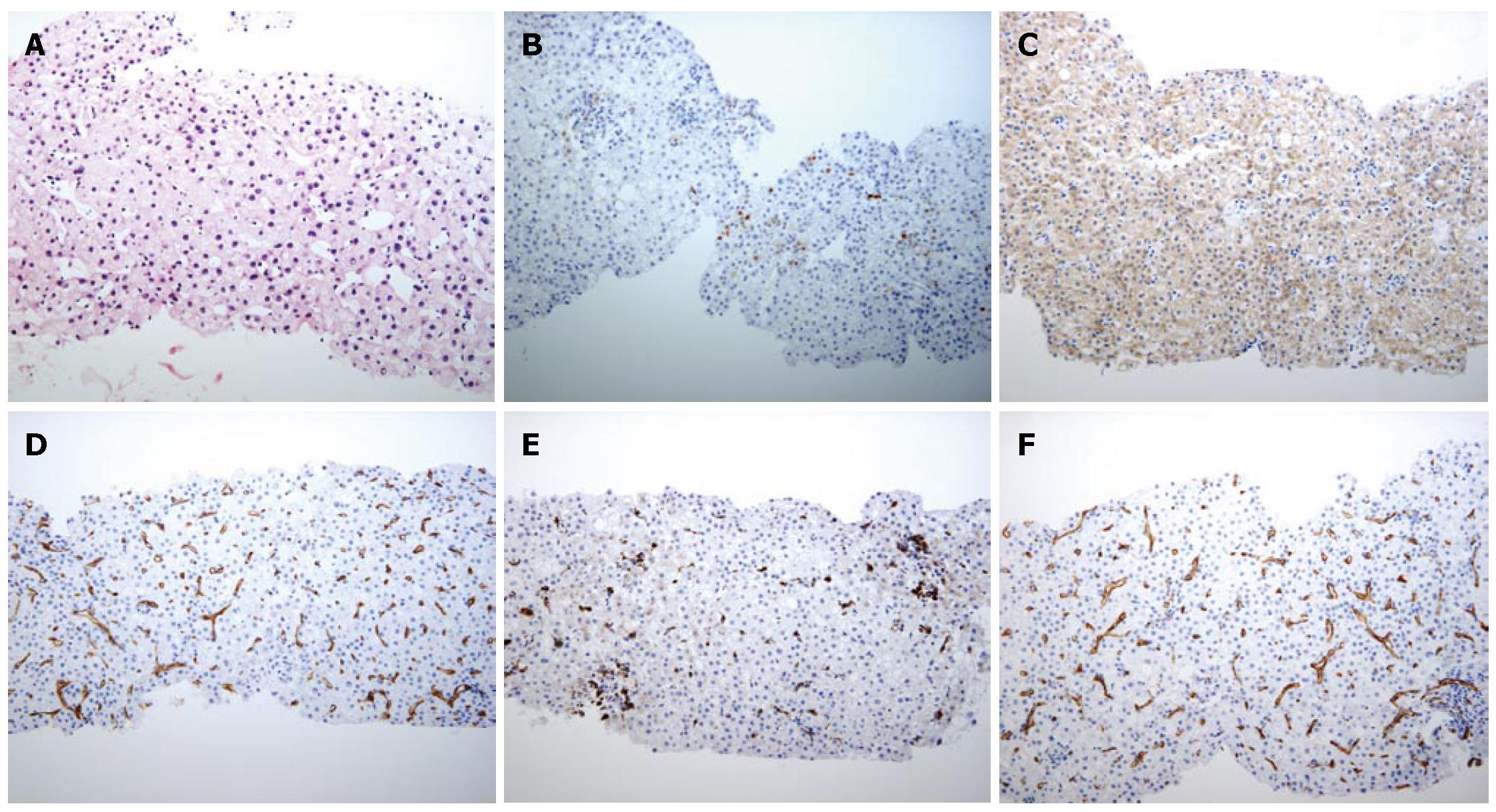
Figure 4 Histological features of a US-guided biopsy of a hyperechoic nodule in S2.
A: Well- to moderately-differentiated HCC characterized by more than two-fold the cellularity of the non-tumorous area, fatty change, clear cell change and mild cell atypia with a thin to mid-trabecular pattern (HE×100); B: Immunohistochemical finding, HSP70 partly positive HCC cells; C: Immunohistochemical finding, CAP2 strongly positive HCC cells; D: Immunohistochemical staining of CD34 in the sinusoidal blood space is positive, showing capillarization; E: Immunohistochemical staining of CD68 Kupffer cells in the sinusoidal blood space is relatively less positive; F: Non-HCC area of immunohistochemical staining of CD68 Kupffer cells in the sinusoidal blood space.
- Citation: Kim SR, Imoto S, Ikawa H, Ando K, Mita K, Fuki S, Sakamoto M, Kanbara Y, Matsuoka T, Kudo M, Hayashi Y. Well to moderately differentiated HCC manifesting hyperattenuation on both CT during arteriography and arterial portography. World J Gastroenterol 2007; 13(43): 5775-5778
- URL: https://www.wjgnet.com/1007-9327/full/v13/i43/5775.htm
- DOI: https://dx.doi.org/10.3748/wjg.v13.i43.5775


