Copyright
©2007 Baishideng Publishing Group Inc.
World J Gastroenterol. Nov 21, 2007; 13(43): 5775-5778
Published online Nov 21, 2007. doi: 10.3748/wjg.v13.i43.5775
Published online Nov 21, 2007. doi: 10.3748/wjg.v13.i43.5775
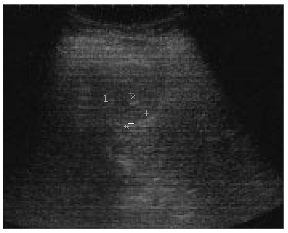
Figure 1 Ultrasound (US) image of a 20-mm hypoechoic nodule in segment 2 (S2).
- Citation: Kim SR, Imoto S, Ikawa H, Ando K, Mita K, Fuki S, Sakamoto M, Kanbara Y, Matsuoka T, Kudo M, Hayashi Y. Well to moderately differentiated HCC manifesting hyperattenuation on both CT during arteriography and arterial portography. World J Gastroenterol 2007; 13(43): 5775-5778
- URL: https://www.wjgnet.com/1007-9327/full/v13/i43/5775.htm
- DOI: https://dx.doi.org/10.3748/wjg.v13.i43.5775


