Copyright
©2007 Baishideng Publishing Group Inc.
World J Gastroenterol. Nov 7, 2007; 13(41): 5537-5539
Published online Nov 7, 2007. doi: 10.3748/wjg.v13.i41.5537
Published online Nov 7, 2007. doi: 10.3748/wjg.v13.i41.5537
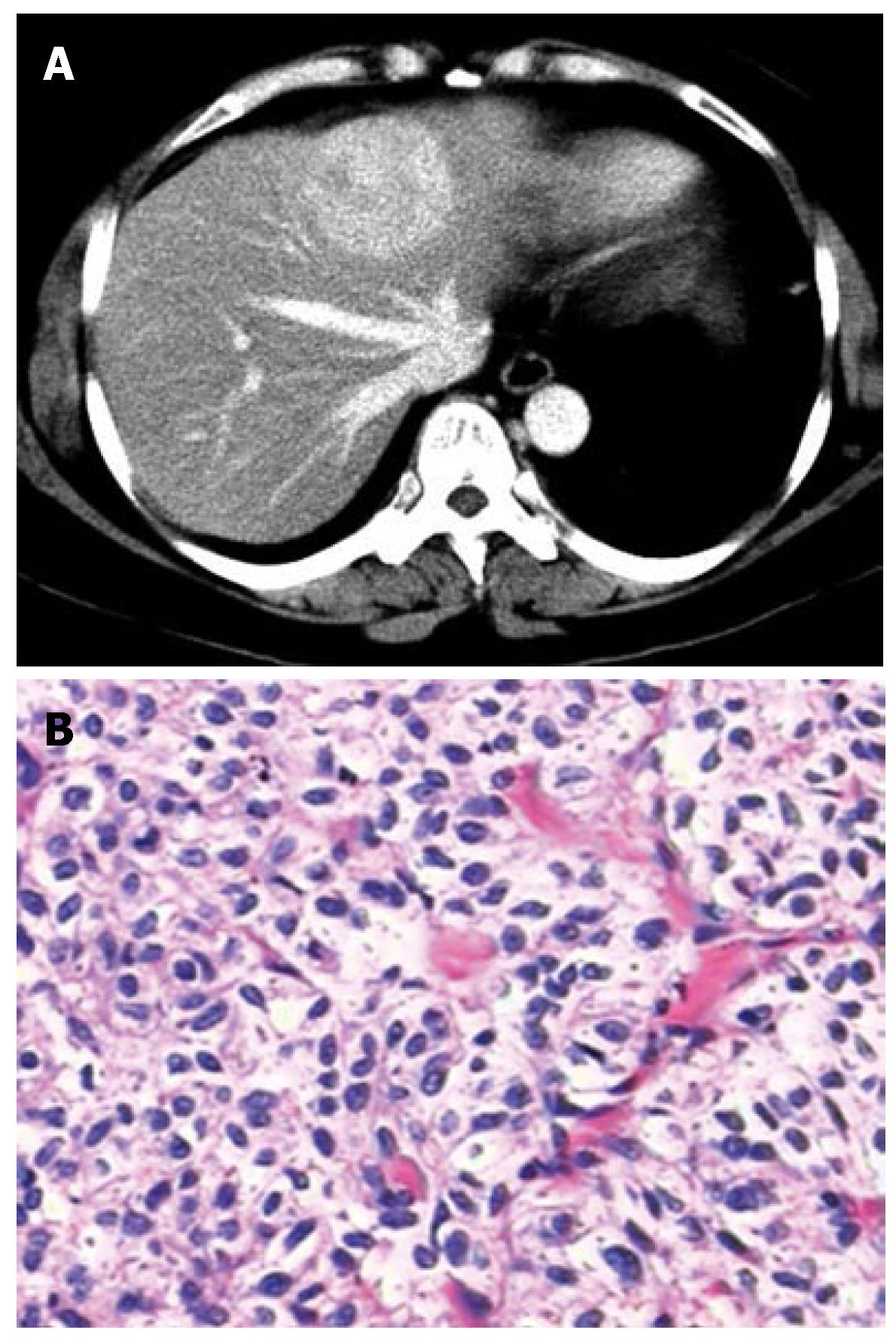
Figure 1 The PEComa of the liver in a 56-year-old woman.
A: Contrast-enhanced CT scan shows significant and heterogeneous enhancement of the lesion; B: Photomicrograph shows polygonal or short spindle cells with oval nuclei and clear abundant cytoplasm (HE, × 200).
- Citation: Fang SH, Zhou LN, Jin M, Hu JB. Perivascular epithelioid cell tumor of the liver: A report of two cases and review of the literature. World J Gastroenterol 2007; 13(41): 5537-5539
- URL: https://www.wjgnet.com/1007-9327/full/v13/i41/5537.htm
- DOI: https://dx.doi.org/10.3748/wjg.v13.i41.5537


