Copyright
©2007 Baishideng Publishing Group Inc.
World J Gastroenterol. Nov 7, 2007; 13(41): 5527-5529
Published online Nov 7, 2007. doi: 10.3748/wjg.v13.i41.5527
Published online Nov 7, 2007. doi: 10.3748/wjg.v13.i41.5527
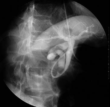
Figure 1 ENBD cholangiogram after endoscopic retrograde cholangiography showing a 1 cm-sized gall stone impacted in a cystic duct joining the extreme lower CBD portion and other stones in gall bladder.
Mild CBD dilation was noted due to the presence of a cystic duct stone.
- Citation: Jung CW, Min BW, Song TJ, Son GS, Lee HS, Kim SJ, Um JW. Mirizzi syndrome in an anomalous cystic duct: A case report. World J Gastroenterol 2007; 13(41): 5527-5529
- URL: https://www.wjgnet.com/1007-9327/full/v13/i41/5527.htm
- DOI: https://dx.doi.org/10.3748/wjg.v13.i41.5527


