Copyright
©2007 Baishideng Publishing Group Inc.
World J Gastroenterol. Nov 7, 2007; 13(41): 5512-5515
Published online Nov 7, 2007. doi: 10.3748/wjg.v13.i41.5512
Published online Nov 7, 2007. doi: 10.3748/wjg.v13.i41.5512
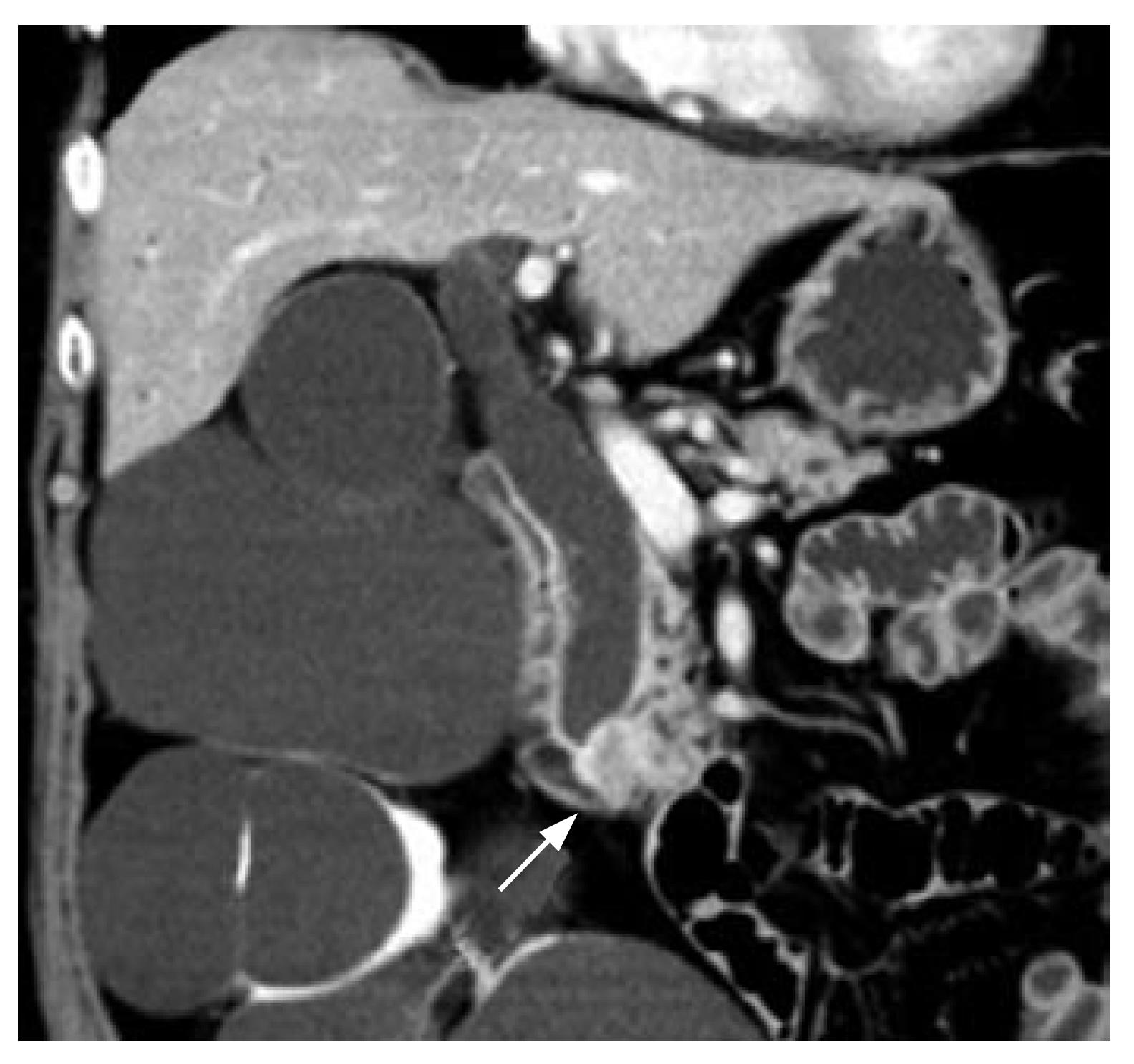
Figure 1 CT reveals a mass in the ampullary region (arrow) with a dilated extrahepatic bile duct.
The patient also had multiple renal cysts.
- Citation: Fujita N, Noda Y, Kobayashi G, Ito K, Obana T, Horaguchi J, Takasawa O, Nakahara K. Histological changes at an endosonography-guided biliary drainage site: A case report. World J Gastroenterol 2007; 13(41): 5512-5515
- URL: https://www.wjgnet.com/1007-9327/full/v13/i41/5512.htm
- DOI: https://dx.doi.org/10.3748/wjg.v13.i41.5512


