Copyright
©2007 Baishideng Publishing Group Inc.
World J Gastroenterol. Oct 28, 2007; 13(40): 5403-5407
Published online Oct 28, 2007. doi: 10.3748/wjg.v13.i40.5403
Published online Oct 28, 2007. doi: 10.3748/wjg.v13.i40.5403
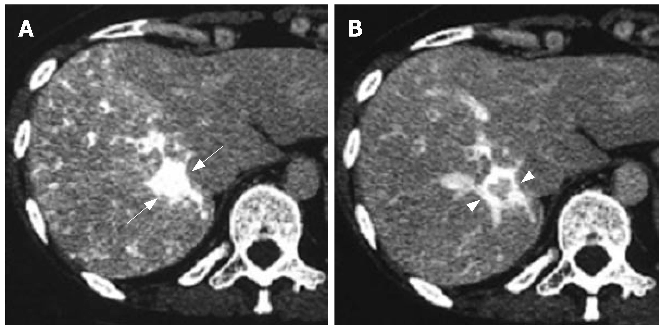
Figure 1 CT angiography showing the conspicuously enhanced lesion in segment 7 through the early phase (arrows) (A) and intensified tumor rim enhancement through the delayed phase along with radial enhancement (arrowheads) (B).
- Citation: Machida T, Takahashi T, Itoh T, Hirayama M, Morita T, Horita S. Reactive lymphoid hyperplasia of the liver: A case report and review of literature. World J Gastroenterol 2007; 13(40): 5403-5407
- URL: https://www.wjgnet.com/1007-9327/full/v13/i40/5403.htm
- DOI: https://dx.doi.org/10.3748/wjg.v13.i40.5403


