Copyright
©2007 Baishideng Publishing Group Inc.
World J Gastroenterol. Oct 28, 2007; 13(40): 5371-5375
Published online Oct 28, 2007. doi: 10.3748/wjg.v13.i40.5371
Published online Oct 28, 2007. doi: 10.3748/wjg.v13.i40.5371
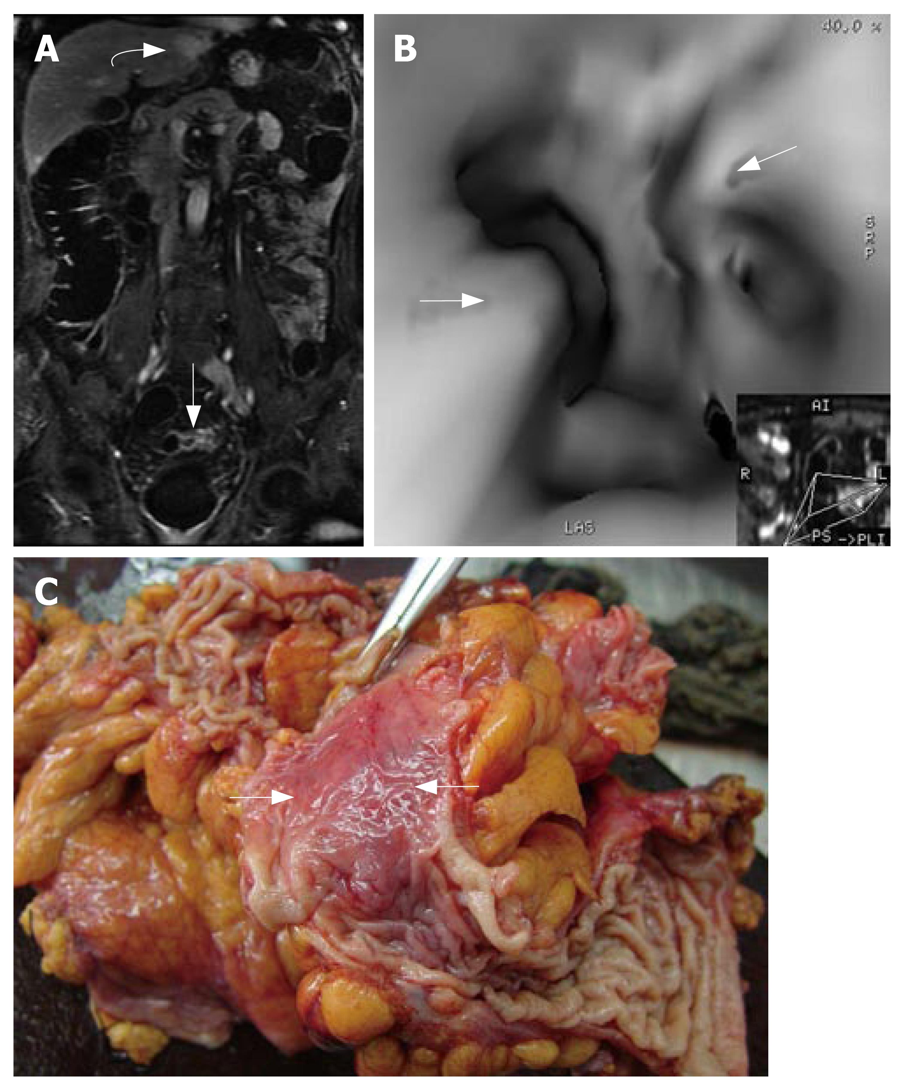
Figure 1 74-year-old man with history of altered bowel habits.
A: The thickened and increased-contrast-uptake bowel wall of sigmoid colon is seen (white arrow) in the coronal gadolinium enhanced T1-weighted image. In liver, an obvious high-intensity neoplasm is discovered (white curved arrow). Subsequent MR scan confirms the diagnosis of a hepatic angioma; B: MR virtual endoscopic rendering confirms stenosis of sigmoid colon (white arrows); C: Post-operation pathology confirms the condition of an inflammatory neoplasm. Specimen shows inflammatory mucosa change (white arrows).
- Citation: Zhang S, Peng JW, Shi QY, Tang F, Zhong MG. Colorectal neoplasm: Magnetic resonance colonography with fat enema-initial clinical experience. World J Gastroenterol 2007; 13(40): 5371-5375
- URL: https://www.wjgnet.com/1007-9327/full/v13/i40/5371.htm
- DOI: https://dx.doi.org/10.3748/wjg.v13.i40.5371


