Copyright
©2007 Baishideng Publishing Group Inc.
World J Gastroenterol. Oct 28, 2007; 13(40): 5351-5356
Published online Oct 28, 2007. doi: 10.3748/wjg.v13.i40.5351
Published online Oct 28, 2007. doi: 10.3748/wjg.v13.i40.5351
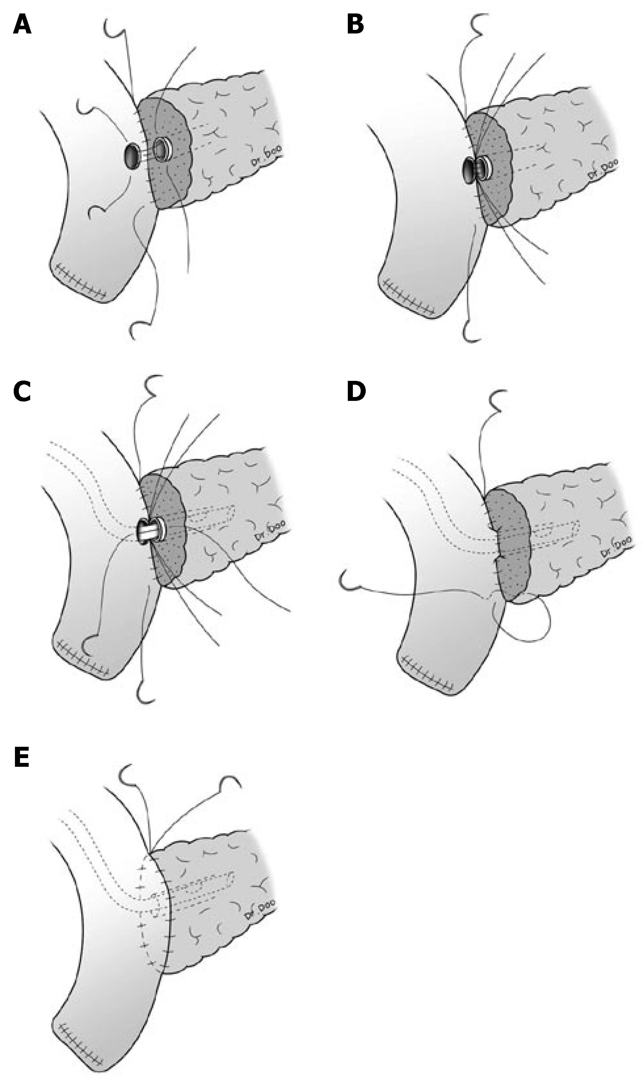
Figure 1 Continuous suture method for the outer layer of pancreaticojejunostomy.
A: The posterior outer layer consisted of the remnant pancreatic parenchyma and the seromuscular layer of jejunum and continuous suture between these two was performed with 5-0 polypropylene (Prolene*, Ethicon, Somerville, NJ); B: The posterior inner layer consisted of the pancreatic duct and mucosa of the jejunum, and interrupted suture for duct-to-mucosa was performed with 5-0 polydioxanone (PDSTMII, Ethicon, Somerville, NJ); C: A silastic polyethylene tube was inserted into the pancreatic duct and external drainage was done; D: For anterior inner layer consisted of the pancreatic duct and mucosa of the jejunum, interrupted suture was performed; E: Continuous suture for anterior outer layer was performed.
-
Citation: Lee SE, Yang SH, Jang JY, Kim SW. Pancreatic fistula after pancreaticoduodenectomy: A comparison between the two pancreaticojejunostomy methods for approximating the pancreatic parenchyma to the jejunal seromuscular layer: Interrupted
vs continuous stitches. World J Gastroenterol 2007; 13(40): 5351-5356 - URL: https://www.wjgnet.com/1007-9327/full/v13/i40/5351.htm
- DOI: https://dx.doi.org/10.3748/wjg.v13.i40.5351


